
Deep Venous Thrombosis — Symptoms, Causes & Treatment
Also known as Deep Venous Thrombosis. Causes, symptoms and how physiotherapy treats Deep Venous Thrombosis — without surgery or medicines.
Understanding Deep Venous Thrombosis
Deep Venous Thrombosis affects people differently, and the right care starts with understanding how it affects you. Its day-to-day impact — on movement, strength, energy and confidence — is where physiotherapy does its work.
Our role in Deep Venous Thrombosis is practical: protect and rebuild movement and daily function with a plan set after a careful assessment, in coordination with your doctor wherever medical management is involved. No two plans look the same, because no two cases behave the same.
Causes & risk factors of Deep Venous Thrombosis
What commonly drives it — your assessment pins down which of these apply to you.
More than one possible driver
Deep Venous Thrombosis can have several contributing factors — a careful history and physical assessment separate them rather than guessing.
Reduced movement and deconditioning
Whatever the trigger, less movement quickly adds weakness and stiffness of its own — usually the most reversible part.
Daily-life load and habits
Work posture, repeated tasks, sleep and stress all shape how a condition behaves day to day.
General health and history
Overall health, previous injuries and existing conditions influence both the picture and the plan — the assessment takes all of it in.
When physiotherapy is the right call
If any of these sound like you, an assessment is the right first step.
- ✓ A new diagnosis of Deep Venous Thrombosis and you want a clear rehabilitation start point
- ✓ Daily tasks, walking or stairs are getting harder than they used to be
- ✓ Strength, stamina or balance has visibly dropped
- ✓ You want a structured clinic + home program with measurable milestones
- ✓ Family members need guidance to support you safely
Your treatment journey
Step by step — from your first assessment to a tracked recovery plan.
A detailed first assessment
History, movement testing and strength checks — a focused session to find what is actually driving your Deep Venous Thrombosis.
A diagnosis you understand
Your physiotherapist explains the findings in plain language — what is affected, why it happened and what that means for you.
Active, tracked treatment
Hands-on therapy, the right modalities and graded exercise — progressed session by session and tracked on Fizo IQ™.
Prevention, built in
The final phase rebuilds strength and habits so the same problem does not come back — with a home plan you keep.
What recovery looks like
- ✓ More independence in daily tasks
- ✓ Better strength, balance and stamina
- ✓ Fewer setbacks, with red flags monitored
- ✓ A program your family understands and can support
- ✓ Progress measured milestone by milestone on Fizo IQ™
When to see a doctor — red flags
How CB Physiotherapy treats Deep Venous Thrombosis
At CB Physiotherapy, treatment for Deep Venous Thrombosis starts with finding its cause: a structured, physiotherapist-led assessment before anything is prescribed. Your plan then combines hands-on therapy, targeted exercise and the right modalities — delivered at a clinic near you or at home, and coordinated with your doctor where medical care is involved.
Every case runs on Fizo IQ™, our recovery engine: diagnosis, root cause, plan, milestones and goals, tracked session by session. You always know where you are in your recovery — and what comes next.
Exercises that help with Deep Venous Thrombosis
Make these exercises yours — a CB physio personalizes, doses and tracks your plan week by week.
Get my personalized planGeneral guidance only — stop if an exercise increases your pain, and check with your physio before starting a new routine.
Get Deep Venous Thrombosis treatment near you
Certified physiotherapists at a clinic near you or at home — same-day slots, assessment-led care.
Request received!
We'll call you within 10 minutes to confirm your home visit.We'll call you within 10 minutes to confirm your slot.
Chat with us on WhatsAppDeep Venous Thrombosis, answered
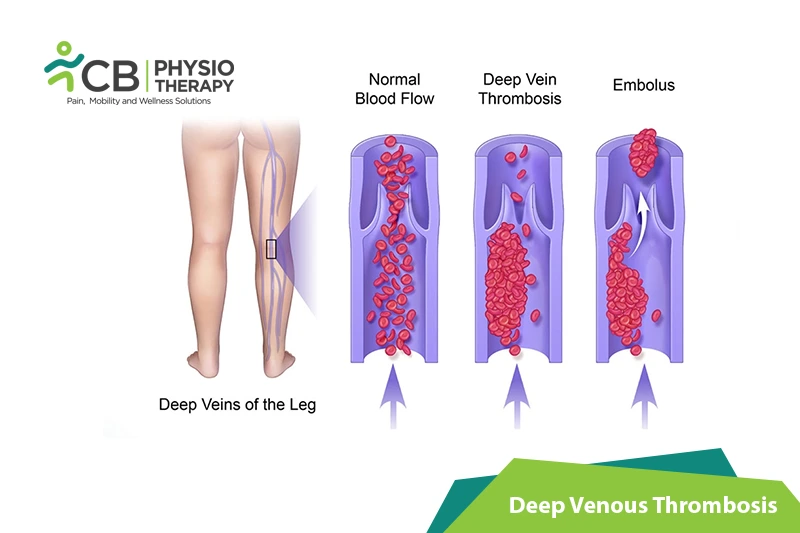
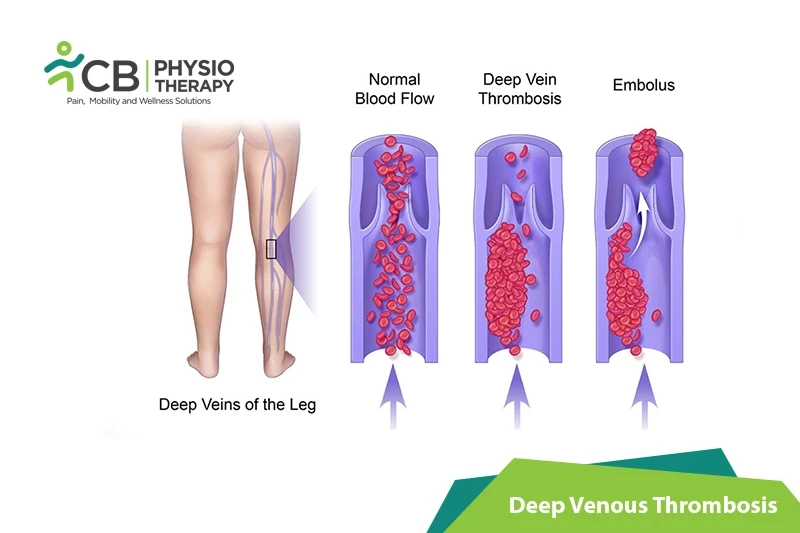
Venous thromboembolism (VTE) is the formation of a blood clot (thrombus) in one or more deep veins that can lead to complications, such as Deep vein thrombosis (DVT), pulmonary embolism (PE), or post-thrombotic syndrome (PTS). Deep vein thrombosis can cause serious complications as the blood clots in the veins can break loose, travel through the bloodstream, and lodge in the lungs, blocking blood flow (pulmonary embolism).
Deep vein thrombosis can occur without symptoms until it happens, but the patient may experience one or more of the following. Deep vein thrombosis symptoms can include:
- Pain often starts in the calf and can be felt like cramping or soreness,
- Swelling in Leg,
- Tenderness in one of the legs usually in the calf,
- Discoloration of skin on the leg,
- Warmth in the affected leg,
- Heavy ache in the affected area
- Red skin, particularly at the back of the leg below the knee
- Difficulty in walking,
- With the progression of the disease, it becomes hard to even stand.
DVT can lead to pulmonary embolism which occurs when a clot blocks one of the blood vessels in the lungs. It can also cause the post-phlebitic syndrome, also known as a post-thrombotic syndrome which damages the veins from the blood clot thus reducing the blood flow in the affected areas.
Pathology:
Deep vein thrombosis is the formation of a blood clot, known as a thrombus, in the deep veins. The deep veins are usually surrounded by powerful muscles that contract and force deoxygenated blood back to the lungs and heart. When the circulation of the blood slows down due to inactivity, illness, or injury, blood can accumulate which provides an ideal setting for clot formation.
Deep venous thrombosis occurs due to a damaged vessel wall, stasis or low flow, hypercoagulability or injury to a vein, or surgery that prevents the blood from circulating or clotting. The causes of thrombosis include:
- Age above 60, have a greater risk of DVT,
- Inheriting a blood-clotting disorder,
- Prolonged bed rest,
- Paralysis,
- Pregnancy,
- Birth control pills (oral contraceptives) or hormone replacement therapy,
- Medical conditions like cancer, heart failure, inflammatory bowel diseases such as Crohn's disease or ulcerative colitis,
- Certain medications and limited movement,
- Overweight or obese,
- Smoking.
Physical examination:
Physical examination is done to check for areas of swelling, tenderness, or discoloration on the skin.
Ultrasound:
A transducer is placed over the part of the body where there's a clot that sends sound waves into the area. These sound waves travel through the tissue and reflect, a computer transforms the waves into a moving image on a video screen and a clot might be visible in the image.
Ultrasonic Imaging:
Series of ultrasounds are done sometimes over several days to determine whether a blood clot is growing or to check for a new one.
Blood test:
People who develop severe deep vein thrombosis usually have an elevated blood level of a substance called D dimer.
Venography:
Dye is injected into a large vein in the foot or ankle and an X-ray creates an image of the veins in the legs and feet, to reveal any blockages.
CT or MRI scans:
CT or MRI scans help to provide visual images of the veins and might also show if there is a clot.
Medication: Blood thinners, Heparin, warfarin or dabigatran, Rivaroxaban, apixaban or edoxaban, clot busters, etc.
Note: Medication should not be taken without the doctor's prescription.
Surgery:
Surgery is done in case the conservative methods do not decrease the symptoms.
Thrombectomy: In a thrombectomy, the doctor injects a contrast dye, which helps to locate the clot using an X-ray. The clot is then removed using a catheter, which is inserted through a vein in the groin or arm and threaded to the location of the blood clot.
Inferior Vena Cava Filter: In this method, medication is used to dissolve large blood clots or placement of an inferior vena cava or filter. In this procedure, a surgeon implants a filter into the inferior vena cava, this filter helps to catch the blood clot that breaks loose from a vein in the legs and is in transit to the lungs, where it can cause a pulmonary embolism. The filter does not treat the blood clot but prevents the clots from traveling to the lungs.
Iliocaval Stenting for Post-Thrombotic Syndrome: DVT can also remain after the treatment. This results in a blockage of the iliac vein in the pelvis and further blockage of blood flow to the heart. It can also damage the valves in the legs that regulate blood flow, allowing blood to flow backward in the veins and collect in the leg. A minimally invasive procedure, iliocaval stenting can provide long-term relief for blockage. In this procedure, an IV is placed into the blood vessel in the groin. The catheter into the blood vessel is delivered. And with the help of continuous X-ray guides small, flexible surgical tools through the catheter and to the site of the blockage. The blockages are opened with stents that act as scaffolding to prop the vein open.
Compression therapy:
Compression stockings help prevent swelling associated with deep vein thrombosis, these are worn on the legs from the feet to about the level of the knees.
Intermittent Pneumatic Compression Device:
Intermittent Pneumatic Compression Device helps to relieve edema and prevent blood clots. It uses cuffs around the legs with air and squeezes the legs. Thus increasing blood flow and preventing blood clots.
Massage therapy helps to stimulate a better response in the affected areas and helps to re-tone the damaged muscle and even suggests special compression techniques that will help the body to recover from the stress caused due to DVT.
Range of motion exercises is done to encourage circulation. e.g
Foot pumps: Place the feet flat on the floor, and then raise the toes towards the body and hold for a few seconds. Lower the toes and balls of the feet to the floor, then raise the heels and hold for another few seconds.
Ankle circles: Raise both the feet off the floor and trace a circle with the toes.
Leg raises: Raise the left foot off the floor. Straighten the leg slowly, and then return the foot to the floor. Repeat with the right leg. Alternatively, lift the knee to the chest, and then bring the foot back to the floor, repeat with the other leg.
Shoulder rolls: Though there are fewer chances to form a clot in the upper body, in case the upper limb is affected, then simply raise the shoulders and circle them back and down 5 times. Then reverse direction for 5 more repetitions.
Strengthening exercises are done with light weights and high repetitions. Strengthening exercises should not be performed after aerobic warm-up.
Stretching Exercises:
Stretching exercises can help to decrease deep venous thrombosis. It decreases muscle soreness and increases flexibility. The patient is recommended to hold the stretch for up to 30 seconds without moving.
Aerobic Exercises:
Walking, swimming, dancing, hiking, and jogging can also help to improve the symptoms of DVT, including, discomfort and swelling.
The patient is educated to avoid sitting still, movement should be done as much as possible. While sitting the patient is advised not to cross the legs, which can hamper blood flow. While traveling a long distance by car, take breaks, stop every hour, and walk around. While on a plane, stand or walk occasionally. If unable to do so then, exercise the lower legs, raise and lower the heels, keep the toes on the floor, then raise the toes with the heels on the floor. Exercise regularly, lose weight and quit smoking to lower the risk of blood clots.
Don't let Deep Venous Thrombosis decide your day.
Get assessed today — find the cause, get a plan, and know when you'll feel better.