Open today · 8 AM–8 PM · Same-day slots
CB Physiotherapy
Same-day appointments

Chondromalacia Patella Treatment Doctors in Ghaziabad

Whether it's a dull ache, a sharp catch, or pain that keeps coming back — we find the root cause of your Chondromalacia Patella and fix it, at a clinic near you or at home.

Precision Care, Proven Recovery
ACCURATE DIAGNOSIS
ROOT CAUSE ASSESSMENT
COMPREHENSIVE RECOVERY PLAN
MILESTONE TRACKING
GOALS ACHIEVED
STAYING ACTIVE
See how it works ↓
Move pain-free Avoid drugs & surgery
5.0 / 5 · 20 Google reviews Verified on Google

Request received!

We'll call you within 10 minutes to confirm your home visit.We'll call you within 10 minutes to confirm your slot.

Chat with us on WhatsApp

Book in 30 seconds

Visit your nearest CB clinic. Tell us your concern and a preferred time — we'll confirm your slot by call within 10 minutes.

Same-day booking by default. We'll confirm your exact slot by call.
+91
No spam · Your number is safe with us
Prefer to talk now? Call +09821966696
Understanding your chondromalacia patella

Does this sound like you?

Common triggers include muscle strain, joint stiffness, poor posture and long desk hours. If two or more of these fit, a physiotherapy assessment can identify exactly what's driving it.

  • A dull ache or sharp, burning chondromalacia patella that keeps returning
  • Discomfort that worsens with movement, lifting or long sitting hours
  • Morning stiffness that eases as you move
  • Pain that limits work, sport or everyday activities
  • Recurring episodes that keep coming back

Most chondromalacia patella improves without surgery or long-term medication — the key is finding the cause first.

⚠ When to seek urgent medical care

Chondromalacia Patella rarely signals something serious, but see a doctor immediately if it comes with:

  • Fever, unexplained weight loss, or a history of cancer
  • Numbness or weakness that keeps getting worse
  • Loss of bladder or bowel control
  • Pain after a major fall or accident

Our physios screen for these red flags at every first assessment and refer you to the right doctor if needed.

Assessment-led treatment

How we treat chondromalacia patella

We start with an AI-based assessment to find the root cause — then follow a clear, four-phase plan.

Phase 1

Ease the pain

Hands-on therapy and advanced modalities to calm pain and inflammation fast.

Phase 2

Restore movement

Mobility work and posture correction so everyday movement feels normal again.

Phase 3

Build strength

Targeted strengthening that protects you — in-clinic or in our physio gym.

Phase 4

Return to activity

Back to work, sport and life — with a home program that prevents relapse.

Your physiotherapist, with Fizo IQ™

Every recovery, planned and proven

One patient record, stage by stage: your physiotherapist pinpoints the diagnosis, finds the root cause and builds your in-clinic plan — while Fizo IQ™, our AI decision-support engine, tracks every milestone until your goals are met and you leave with an exercise plan to stay active.

Physio Expert @CB with Fizo IQ™ — recovery engine
TRACKING
ACCURATE DIAGNOSIS
L4–L5 disc bulge · nerve-root irritation, pinpointed at the first movement assessment
ROOT CAUSE ASSESSMENT
Poor sitting posture, weak core–hip chain · quietly loading that disc
COMPREHENSIVE RECOVERY PLAN
24 clinic sessions · manual therapy + spinal decompression · guided exercise therapy, reviewed weekly
MILESTONE TRACKING
Sitting pain-free ✓ · stairs ✓ · next: the morning 5 km walk
GOALS ACHIEVED
Pain 7 → 3 in six weeks · back to daily life, moving freely
STAYING ACTIVE
A personalized exercise plan, yours to keep · so the pain doesn't come back
Example recovery journey · every plan is built around you
Feeling something?

Symptoms Chondromalacia Patella addresses

These symptoms respond well to Chondromalacia Patella — tap one to see how we treat it near you.

Move better

Exercises that help with Chondromalacia Patella

More exercises →

Make these exercises yours

Get my personalized plan

General guidance only — stop if an exercise increases your pain, and check with your physio before starting a new routine.

Meet your care team

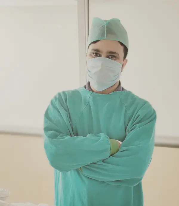
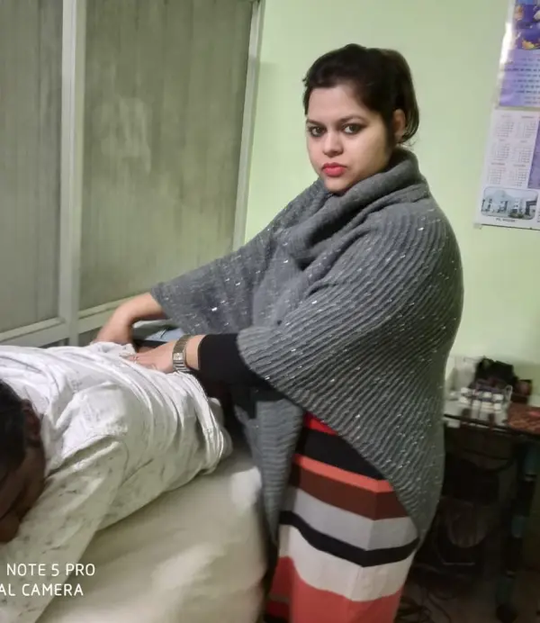
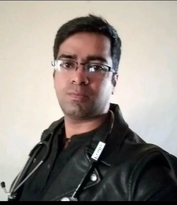
Licensed experts who treat hundreds like you every month

Every CB physio is a qualified BPT/MPT clinician. You don't need to choose one — our lead physiotherapist reviews your case and assigns the specialist best suited to treat it.

Dr. Puneet Kumar, physiotherapist in Ghaziabad Verified

Dr. Puneet Kumar

Physiotherapist · MPT (Orthopaedics)

5.0 (1)
Ghaziabad
6 yrs experience
Ortho Spine Sports Physiotherapy Center
Not sure who to see? You don't have to decide. Our lead physiotherapist reviews every case and assigns the physio best suited to it — based on your condition and availability.
Patient stories

Chondromalacia Patella recoveries, verified on Google

Rated 5.0 / 5 across 20 verified patient reviews in Ghaziabad.

Verified

“मुझे फिजियोथेरेपी करवानी है होम सर्विस आपके पास अवेलेबल है”

SK
Sumit Kumar Verified patient · Treated at CB · 5 months ago
Verified

“VERY Best physiotherapist and good experience”

AU
Arun Upadhyay Verified patient · Treated at CB
Verified

“I recently had the pleasure of receiving physiotherapy treatment from Dr. Asif, and I cannot praise his skills and dedication enough. As a physiotherapist, he possesses a unique blend of expertise, compassion, and professionalism that sets him apart from others in his field.”

SK
Shawaiz Khan Verified patient · Treated at CB · 3 months ago
Verified

“I recently had the privilege of being treated by Dr. Asif Alam, and I cannot speak highly enough of his exceptional care and expertise as a physiotherapist. His ability to diagnose and explain the root causes of my issues was truly impressive. He provided me with a clear understanding of my condition His hands-on techniques were precise and effective, providing me with immediate relief and tangible results. He continuously monitored my progress and adjusted the treatment plan accordingly, ensuring that I was on track to a full recovery.”

KN
Krishna N12 Verified patient · Treated at CB · 3 months ago
Verified

“I have no words to express how good Dr. Asif was for post-knee rehabilitation. Every session I had was excellent. Day by day, I'm improving. I can walk without support. Thanks so much! All praise for you!🙏”

RB
Rajiv Bhatia Verified patient · Treated at CB · 3 months ago
Verified

“Thank you, Dr. shally for your outstanding service!👍🏼”

AB
Aakash Bansal Verified patient · Treated at CB · 4 months ago
Good to know

Chondromalacia Patella, answered

Chondromalacia patella or anterior knee pain or runner's knee is the softening and breakdown of the hyaline cartilage on the underside of the kneecap or the patella, resulting in pain when the knee and the thigh bone rub with each other. Most commonly it involves the extensor mechanism of the knee.

The most common symptom of CMP is anterior knee pain, which aggravates due to the activities that load the patellofemoral joint such as running, stair climbing, squatting, kneeling, or changing from standing to a sitting position. Other symptoms include:

  • Tenderness on the medial and lateral side of the patella,
  • Crepitations felt on motion,
  • Swelling,
  • Weakness or tightness of the muscles associated with the knee,
  • Weakness in vastus medialis muscle and a high Q angle,
  • Feeling of grinding when the knee is flexed may occur E.g. while bending knees, going downstairs, running downhill, and standing up after sitting for a while.

Pathology:

Chondromalacia or sick cartilage is an affliction of the hyaline cartilage coating of the articular surfaces of the bone. It occurs due to the softening and then subsequent tearing, and erosion of hyaline cartilage. It commonly involves the extensor mechanism of the knee and is often referred to as chondromalacia of the patella, patellofemoral syndrome, or runner's knee. The undersurface of the patella is covered with hyaline cartilage that articulates with the hyaline cartilage-covered femoral groove or trochlear groove. Microtrauma wear and tear, post-traumatic injuries, and certain injections of medication can lead to chondromalacia. Chondromalacia occurs in any joint and is most common in joints that have had trauma and deformities. Repeated activities and wear and tear create compressive stress on the patella-femoral joint or increased loads applied to the joint can lead to chondromalacia.

Chondromalacia patella occurs when there is continuous rubbing together of the knee and the thigh bone due to wear and tear. This causes aching pain or a feeling of grinding when the knee is flexed. The causes of chondromalacia can be:

  • Injury,
  • Generalized constitutional disturbance and patellofemoral contact,
  • Trauma to chondrocytes in articular cartilage,
  • Instability or malt racking of patellae,
  • Malalignment of the femur,
  • Excessive activity involving the knee,
  • Flat feet,
  • Overweight,
  • Dislocation or fracture related to the kneecap,
  • Sportspersons like bicyclists, runners, soccer players, and other people who exercise often.

Physical examination:

Physical examination of the knee is done to determine the cause of pain. Usually, joint appearance is normal but there appears to be slight effusion, passive movements are usually painless and free, but the repeated extension of the knee from flexion produces pain and a grating-like feeling under the patella, especially if the articular surfaces are compressed together. Pain and crepitus are felt if the patella is compressed against the femur, with the knee in full extension. Sharp pain under the patella is felt on resisting a static quadriceps contraction.

Clark test:

This test is done to detect patellofemoral joint disorder. The patient is in a supine position with the knee extended, the examiner then places the webspace of his hand superior to the patella while applying pressure. The patient is instructed to gently contract the quadriceps muscle. The test is positive if the patient feels is a pain in the patellofemoral joint.

Patellar tap test

The patient is in a supine position with the legs extended. The examiner applies pressure on the proximal side of the knee to squeeze out the fluid from the suprapatellar pouch. The fluid can be moved under the patella while maintaining the pressure on the suprapatellar pouch, the examiner uses the hand to press upon the medial and lateral recesses forcing the fluid under the patella. The therapist then taps down the patella with the index finger to create an upward and downward movement, on doing so when the test is a positive sign the patella floats or bounces and indicates knee joint effusion.

X-ray:

Though in most of the cases there are no radiological changes found, but in the later stages, patellofemoral joint space narrows and osteoarthritic changes begin to appear. X-ray also helps to rule out some types of arthritis or inflammation.

MRI and CT scan:

Magnetic resonance imaging (MRI) and CT scan shows the details of the knee joint and reveals many cases of chondromalacia patella. Detailed images of the soft tissue and bony structures of the knee joint are seen in more detail than X-rays.

Arthroscopy:

In the arthroscopy method tiny, flexible camera is inserted into the knee to see the image of the cartilage looks.

Medication: Nonsteroidal anti-inflammatory drugs (NSAIDs), topical pain medication, analgesics, etc.

Note: Medication should not be taken without the doctor's prescription.

Surgery:

In case conservative measures fail to reduce the symptoms then there are several possible surgical procedures such as:

  • Chondrectomy.
  • Partial removal of the patella.
  • Full patellectomy.
  • Replacement of the damaged cartilage.
  • Drilling is used to heal damaged cartilage.
  • Tightening of the medial capsule (MC) (in case of the MC is lax).
  • Lateral release (in case of the very tight lateral capsule will pull the patella laterally).
  • The medial shift of the tibial tubercle.

Rest:

The most common way to treat symptoms of chondromalacia patella is to immobilize the knee.

Braces:

Bracing the patella and the knee joint helps to reduce pain and symptoms, but alters patella tracking and reduces the active function of the quadriceps. Bracing may be useful to offer patients some support and pain relief in the short term and also helps to avoid antalgic movements and normalize gait as much as possible. The brace should allow variation in a medial pull on the patellar and pressure and thus can be used for patient’s pre-and postoperatively. Patellar realignment brace can also be used.

Foot Orthoses:

Foot orthoses also help for pain relief, but in cases where lower limb mechanics are deemed to be contributing to the knee pain, which may be due to excessive lower limb internal rotation during weight-bearing, poor pronation control, and increased Q-angle.

Cold therapy:

Ice therapy may be used for reducing pain in an acute stage but not in long-term treatment. It is used to relieve inflammation and reduce pain. Ice massage is done by applying ice to the area of inflammation, it can be used as a cold pack for 5-7 minutes, but care should be taken to avoid frostbite.

Transcutaneous electrical nerve stimulation (TENS):

Transcutaneous electrical nerve stimulation (TENS) is an electrotherapy modality that provides pain relief by pain modulation. Transcutaneous electrical nerve stimulation closes the gate mechanism at the anterior grey horn in the spinal cord and stimulates the endogenous opioid system which prevents the release of substance p at the anterior grey horn.

Short wave diathermy (SWD):

Short wave diathermy is a deep heating modality that uses heat to provide pain relief, it improves the blood supply to the muscles and removes waste products.

Interferential current therapy (IFT):

Interferential current therapy produces a low-frequency effect on the tissue. It inhibits the transmission of pain impulses and stimulates the endogenous opioid system, increases blood supply, relieves edema, and also removes waste products.

Kinesio- taping

Kinesio-Taping of the patella is done to influence its movement to provide some short-term relief.

Exercises:

Exercises like gentle Isometric quadriceps exercise, relaxed passive or active knee swinging to maintain a full range of motion, single-leg raise, strong hip ankle-foot movements.

Strengthening and stretching exercises such as hamstring stretching exercise, hip strength, and stability training, hip abductor strengthening exercise program, stretching exercise for rectus femori, gastrocnemius, and tensor fascia lata, strengthening exercise of vastus medialis oblique muscle.

Foam roller:

The foam roller is used to relieve tight musculature and reduce pressure over the patella such as foam roller exercise for hamstring muscle, gastrocnemius muscle, iliotibial band, and tensor fascia lata.

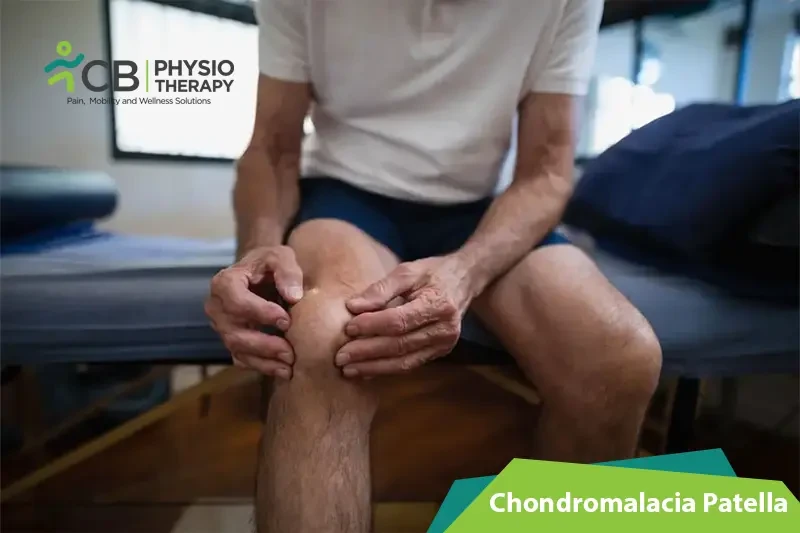
The patient is advised to avoid strenuous activities until the pain eases. Symptoms usually improve in the set time if the knee is not overused. Patient should also be advised to lose weight and wear the right kind of sports or running shoes and use special shoe inserts and support devices. The patient should be made aware that long-term recovery occurs in teenage patients because their bones are still growing and symptoms tend to disappear once adulthood is reached.

Ready when you are

Don't let the pain decide your day.

Get assessed today — find the cause, get a plan, and know when you'll feel better.

📞 Call 💬 WhatsApp Book now