Verified
Dr. Deepanshu Khatri
Physiotherapist
See clinics, physiotherapists and prices for your city.
Whether it's a dull ache, a sharp catch, or pain that keeps coming back — we find the root cause of your Meniscal Injury and fix it, at a clinic near you or at home.
Precision Care, Proven RecoveryWe'll call you within 10 minutes to confirm your home visit.We'll call you within 10 minutes to confirm your slot.
Chat with us on WhatsAppCommon triggers include muscle strain, joint stiffness, poor posture and long desk hours. If two or more of these fit, a physiotherapy assessment can identify exactly what's driving it.
Most meniscal injury improves without surgery or long-term medication — the key is finding the cause first.
Meniscal Injury rarely signals something serious, but see a doctor immediately if it comes with:
Our physios screen for these red flags at every first assessment and refer you to the right doctor if needed.
We start with an AI-based assessment to find the root cause — then follow a clear, four-phase plan.
Hands-on therapy and advanced modalities to calm pain and inflammation fast.
Mobility work and posture correction so everyday movement feels normal again.
Targeted strengthening that protects you — in-clinic or in our physio gym.
Back to work, sport and life — with a home program that prevents relapse.
One patient record, stage by stage: your physiotherapist pinpoints the diagnosis, finds the root cause and builds your in-clinic plan — while Fizo IQ™, our AI decision-support engine, tracks every milestone until your goals are met and you leave with an exercise plan to stay active.
Every plan combines hands-on care, advanced modalities and guided exercise — matched to your assessment.
Make these exercises yours — a CB physio personalizes, doses and tracks your plan week by week.
Get my personalized planGeneral guidance only — stop if an exercise increases your pain, and check with your physio before starting a new routine.
Every CB physio is a qualified BPT/MPT clinician. You don't need to choose one — our lead physiotherapist reviews your case and assigns the specialist best suited to treat it.
Verified
Physiotherapist
Head Of The Clinic
Head Of The Clinic · BPT, MPT
Verified
Sr. Consultant Physiotherapist · BPT, MPT
Head Of The Clinic · BPTh, MPTh
Sr. Consultant Physiotherapist · BPTh
Head Of The Clinic · BPTh, MPTh
The same prices you'll see at the clinic — published before you book. Sessions run 30–45 minutes.
Booking a course of sessions? Save 10% when you take a treatment package — and recover with a plan built around your goals.
Ask about packages →Rated 5.0 / 5 across 102 verified patient reviews in Delhi.
“I visited CB Physiotherapy because I pulled my hamstring. I consulted Dr. Meghna here who gave me a one week treatment plan and suffice to say I started to see results after the first session itself. I was unable to sit and had a lot of pain while walking. After my first session, I was able to walk better and sit better. By the end of the week, I had no pain. They have good infrastructure here and offer various treatments from cupping therapy to laser treatment. I would definitely recommend Dr. Meghna over any other physiotherapist in Saket if not Delhi.”
“I went for consultation for my knee pain and there I met Dr Rahul He took my consultation and sessions for 9 days and he was supr throughout the sessions and now it's so easy for me to be back in daily routine.”
“I had an amazing experience with Dr. Rahul also he took care of everything regarding my lower back strain also they have all the equipment which can help them to cure from the pain or injury. It was a great overall experience with the clinic staff too”
“Hello, I had 15 sessions with them. Overall all sessions were good. Their staff are very knowledgeable and helpful. A special thanks to Mr. Rahul and Ms. Mehak. Both them really helped with my pain on right shoulder and explained about basic and various exercises which i should do on regular basis. Would definitely recommend others.”
“Dr. Deepanshu is an exceptional physiotherapist! His personalized treatment plan and hands-on techniques helped me recover from chronic back pain effectively. He’s patient, knowledgeable, and takes the time to explain everything clearly. His positive attitude and encouragement made the entire process motivating and stress-free. I highly recommend him to anyone seeking top-notch physiotherapy care!”
“Dr. Rahul is an amazing physiotherapist! He genuinely cares about his patients and tailors treatments to fit your needs. Thanks to him, I’ve seen great progress and feel so much better. Highly recommend.”
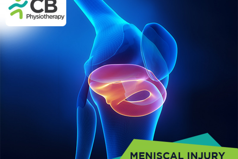
The semilunar cartilages are commonly called menisci and form an important shock-absorbing mechanism, which helps in the smooth functioning of the knee. They help in the gliding movement of the tibia on the femur.
The menisci are prone to injuries due to overload and overstretching. The other causes may be:
· Poor strength
· Poor flexibility
· Fatigue
· Imbalance
· Previous injury
· Overuse
These injuries are most commonly seen in:
· Athletes involved in soccer, football, kabaddi.
There are two menisci in a knee the medial menisci and the lateral menisci. The symptoms can be seen in anyone or both the menisci.
· Pain
· Tenderness
· Swelling
· Inflammation
· Locking of the knee
· Clicking sound
· Loss of knee movement
Pathology
There are three types of meniscal tears, longitudinal tear, horizontal tear, and oblique tear. Further longitudinal subtypes are bucket handle tear, posterior tear, and anterior tear.
Abduction external rotation violence, on the flexed weight-bearing knee, causes a tear in the medial meniscus. During football, it occurs when the player, standing on one leg which is slightly flexed at the knee, turns to tackle the ball with the other leg.
The lateral meniscus is damaged by the opposite violence i.e. internal rotation and abduction violence of the tibia on a semi-flexed weight-bearing knee.
Magnetic resonance imaging (MRI)
On Magnetic resonance imaging (MRI) acute injuries typically show up as the high signal intensity.
Plain radiographs
Plain radiographs should be obtained as the initial imaging modality to rule out a fracture, and evaluate any avulsion injuries.
Ultrasound
Ultrasound maybe used as a secondary tool to evaluate partial or complete tears
Special clinical tests
McMurray's test:
This test elicits a click of the cartilage if it is torn.
Apley's grinding test:
When the meniscus is torn, pain, and click localized to the joint line are elicited.
Bounce home test for bucket handle type tear:
If the extension is not complete or has a rubbery end-feel with elastic resistance to blocking full extension, it indicates bucket handle type of meniscal tear.
Oral non-steroidal anti-inflammatory drugs (NSAIDs) like naproxen, ibuprofen. Corticosteroid injection
(Medication not to be taken without doctor's prescription).
Techniques used by the physiotherapist:
· NMES:
Neuromuscular stimulations facilitate voluntary motor control and muscle strength.
Application of ultrasound decreases pain, increases range of motion, and decreases inflammation.
· Rest:
Short immobilization is advantageous in limiting the extent of damage at the site of the injury. Use of crutches and bed rest is important.
The physiological effects of ice are beneficial in the healing process should be applied in a plastic bag and wrapped directly over the affected area.
· Compression:
Gentle compression is used with a firm compressive bandage placed around the knee.
· Elevation:
Protection of the knee from the position of stress. It helps to reduce edema and allows the return of fluid to the heart, elevate the extremity above the heart level.
· Isometric exercises:
Isometric exercises for the quadriceps. These should be done at least for 5 minutes every hour. Prevention of quadriceps and reflex inhibition is important. The isometrics progressed slowly to the maximum and sustained for 10 -15 seconds, assist in strengthening the quadriceps, whereas speedy isometrics help in the resolution of edema and effusion.
· Ankle-toe movements:
When the leg is immobilized in a pop cast, vigorous movements to the ankle and toes are important to prevent venous thrombosis and to augment circulation.
· Heel slides:
Sit on a firm surface with a socks on the foot or a towel under the heel. Gently flex the knee with the foot approaching the buttocks, then return to the starting position.
· Wall slides:
Lie on a firm surface with the feet resting on the wall. Slowly begin to walk the feet down the wall.
· Straight leg raise (SLR):
SLR, straight leg raise should be initiated as an assisted movement. It can be made more effective by, slow repetitions with self-generated intramuscular tension without the heel making contact with the bed.
· Straight leg abduction (SLR modified to SLA):
SLA, straight leg abduction exercise has advantages of the two movements are that all-important muscle groups of the lower limb are exercised at the same time. Due to sustained contractions in all muscle groups, strength, as well as endurance, also improves to a great extent.
· Knee swinging:
Relaxed knee swinging should be made speedy with increased arc of movement to attain free mobility.
· Progressive resistive exercises(PRE):
Gradual self-resistive exercise with self-generated tension or graded resistance exercise with weight belts.
· Flexibility exercises:
Flexibility exercises of the static stretching using a 30-the second hold, relaxing, and performing 5 repetitions to hamstrings, iliotibial tract, and gastrocsoleus are important.
· Modified Straight leg raise (SLR):
By adding weight, the weight or resistance should be gradually progressive. The upper limit of resistance for the SLR should not be kept at 4.5 to 6.8kg. (as permitted by pain and achieved ROM, so as not to overstrain the knee.)
· Knee flexion:
Knee flexion exercises should be practiced in prone as well as on a static bicycle. Speed resistance and the seat height of the static bicycle should be properly adjusted so as not to overstrain the knee. Begin with a half-circle of the peddle. The session should be for 15 minutes initially, increasing gradually to an hour.
· Standing concentric isotonic exercises:
Stand near a table or wall for support. Place a cuff weight on the involved leg to bring the heel toward the buttocks in a slow controlled manner.
· Vigorous program:
When the pain is minimal, ROM and swelling are near normal. The endurance strengthening flexibility exercises are made progressive by suitable techniques. Progress to guided prone kneeling, assisted squatting, stair climbing and descending and cross leg sitting.
· Isokinetic exercises:
Isokinetic mode of exercise is effective in improving strength endurance and functional capacity of the quadriceps and hamstrings. It assists in improving ROM, due to the physiological overflow. As resistance is adjusted in relation to the muscle force through ROM, it prevents excessive compression of the joint. As the isokinetic provides fast contractile velocity, it improves control and coordination.
Aerobic exercises like running, jogging, and swimming can be initiated as the pain permits.

Get assessed today — find the cause, get a plan, and know when you'll feel better.