Open today · 8 AM–8 PM · Same-day slots
CB Physiotherapy
Same-day appointments

Medial Collateral Ligament (mcl) Injury Treatment Doctors in Gurgaon

Whether it's a dull ache, a sharp catch, or pain that keeps coming back — we find the root cause of your Medial Collateral Ligament (mcl) Injury and fix it, at a clinic near you or at home.

Precision Care, Proven Recovery
ACCURATE DIAGNOSIS
ROOT CAUSE ASSESSMENT
COMPREHENSIVE RECOVERY PLAN
MILESTONE TRACKING
GOALS ACHIEVED
STAYING ACTIVE
See how it works ↓
Move pain-free Avoid drugs & surgery
5.0 / 5 · 22 Google reviews Verified on Google

Request received!

We'll call you within 10 minutes to confirm your home visit.We'll call you within 10 minutes to confirm your slot.

Chat with us on WhatsApp

Book in 30 seconds

Visit your nearest CB clinic. Tell us your concern and a preferred time — we'll confirm your slot by call within 10 minutes.

Same-day booking by default. We'll confirm your exact slot by call.
+91
No spam · Your number is safe with us
Prefer to talk now? Call +09821966696
Understanding your medial collateral ligament (mcl) injury

Does this sound like you?

Common triggers include muscle strain, joint stiffness, poor posture and long desk hours. If two or more of these fit, a physiotherapy assessment can identify exactly what's driving it.

  • A dull ache or sharp, burning medial collateral ligament (mcl) injury that keeps returning
  • Discomfort that worsens with movement, lifting or long sitting hours
  • Morning stiffness that eases as you move
  • Pain that limits work, sport or everyday activities
  • Recurring episodes that keep coming back

Most medial collateral ligament (mcl) injury improves without surgery or long-term medication — the key is finding the cause first.

⚠ When to seek urgent medical care

Medial Collateral Ligament (mcl) Injury rarely signals something serious, but see a doctor immediately if it comes with:

  • Fever, unexplained weight loss, or a history of cancer
  • Numbness or weakness that keeps getting worse
  • Loss of bladder or bowel control
  • Pain after a major fall or accident

Our physios screen for these red flags at every first assessment and refer you to the right doctor if needed.

Assessment-led treatment

How we treat medial collateral ligament (mcl) injury

We start with an AI-based assessment to find the root cause — then follow a clear, four-phase plan.

Phase 1

Ease the pain

Hands-on therapy and advanced modalities to calm pain and inflammation fast.

Phase 2

Restore movement

Mobility work and posture correction so everyday movement feels normal again.

Phase 3

Build strength

Targeted strengthening that protects you — in-clinic or in our physio gym.

Phase 4

Return to activity

Back to work, sport and life — with a home program that prevents relapse.

Your physiotherapist, with Fizo IQ™

Every recovery, planned and proven

One patient record, stage by stage: your physiotherapist pinpoints the diagnosis, finds the root cause and builds your in-clinic plan — while Fizo IQ™, our AI decision-support engine, tracks every milestone until your goals are met and you leave with an exercise plan to stay active.

Physio Expert @CB with Fizo IQ™ — recovery engine
TRACKING
ACCURATE DIAGNOSIS
L4–L5 disc bulge · nerve-root irritation, pinpointed at the first movement assessment
ROOT CAUSE ASSESSMENT
Poor sitting posture, weak core–hip chain · quietly loading that disc
COMPREHENSIVE RECOVERY PLAN
24 clinic sessions · manual therapy + spinal decompression · guided exercise therapy, reviewed weekly
MILESTONE TRACKING
Sitting pain-free ✓ · stairs ✓ · next: the morning 5 km walk
GOALS ACHIEVED
Pain 7 → 3 in six weeks · back to daily life, moving freely
STAYING ACTIVE
A personalized exercise plan, yours to keep · so the pain doesn't come back
Example recovery journey · every plan is built around you
Feeling something?

Symptoms Medial Collateral Ligament (mcl) Injury addresses

These symptoms respond well to Medial Collateral Ligament (mcl) Injury — tap one to see how we treat it near you.

Move better

Exercises that help with Medial Collateral Ligament (mcl) Injury

More exercises →

Make these exercises yours

Get my personalized plan

General guidance only — stop if an exercise increases your pain, and check with your physio before starting a new routine.

Meet your care team

Licensed experts who treat hundreds like you every month

Every CB physio is a qualified BPT/MPT clinician. You don't need to choose one — our lead physiotherapist reviews your case and assigns the specialist best suited to treat it.

Not sure who to see? You don't have to decide. Our lead physiotherapist reviews every case and assigns the physio best suited to it — based on your condition and availability.
Patient stories

Medial Collateral Ligament (mcl) Injury recoveries, verified on Google

Rated 5.0 / 5 across 22 verified patient reviews in Gurgaon.

Verified

“Best physiotherapist on CB physio platform . Highly educated and very confident regarding knowledge. Thanks CB physio you provide me best Doctor...”

JB
Jatin Verified patient · Treated at CB
Verified

“Received good treatment from Dr .Amit for ankle injury. Now I can walk without any support. My mobility is improved so much..🙏🙏 thank you”

AM
Aman Mehra Verified patient · Treated at CB · 3 months ago
Verified

“Thanks for the treatment, sir, for my back issue; it is quite relaxing now. Pain has been subsidised a lot. I am very satisfied with the treatment I received.”

PS
Prajakta Sharma Verified patient · Treated at CB · 3 months ago
Verified

“i have been to several physical therapy providers over the years, and this one is by far the best. The therapists is caring and really know their stuff. i got treatment for elbow at my home. its so convenient. the treatment was good i can feel much difference..thank you.”

AA
Ashish Arora Verified patient · Treated at CB · 3 months ago
Verified

“I suffered a serious injury that left me with limited mobility and a lot of pain. But thanks to the amazing care I received from Dr. Amit, I was able to regain my strength after 4 sessions and get back to my normal routine. I’m so grateful for his support and expertise..”

GB
Gautam Brar Verified patient · Treated at CB · 3 months ago
Verified

“Dr. Surender is an exceptional home physiotherapy services provider who has a wealth of knowledge and experience in treating various conditions. I recently had the pleasure of receiving treatment from him for a persistent back pain, and I can confidently say that he is one of the best physiotherapists I have ever had.👍🏼🙏”

AS
Amritpal Singh Verified patient · Treated at CB · 4 months ago
Good to know

Medial Collateral Ligament (mcl) Injury, answered

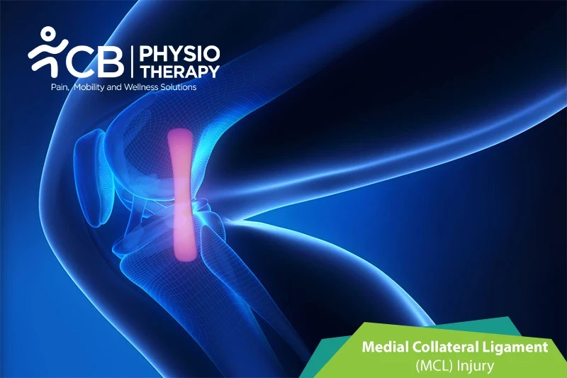
Knee joint allows the lower leg to bend and straighten, the movement is controlled and protected by the ligaments and the muscles in the knee. Ligaments are tough bands of tissue that attach to the bones on each side. There are four types of ligaments in the knee, these are:

  • The medial collateral ligament (MCL), present on the medial aspect of the knee or inside of the knee.
  • The lateral collateral ligament (LCL), present on the lateral aspect of the knee or outside of the knee.
  • The anterior cruciate ligament (ACL) and posterior cruciate ligament (PCL), prevent the frontward or anterior and backward posterior movement of the knee joint.

The MCL is located on the medial aspect of the knee and is attached to the thighbone and shinbone. The MCL holds the knee stable during valgus stress that is when stress is put on the outer part of the leg that can buckle the knee toward the center of the body.

Knee injuries that involve the MCL injuries are referred to as sprains or tears.

MCL sprains can be graded as follows:

  • Grade I (fibers are stretched).
  • Grade II (ligament fibers are partially torn).
  • Grade III ligament is completely torn).

MCL is usually related to the Medial meniscus and the anterior cruciate ligament (ACL), these two structures may also be damaged in association with an MCL injury.

Medial collateral ligament injuries are the most common ligament injuries of the knee. The reasons for the injury can be:

  • Males are at more risk than females,
  • Sports injuries like contact sports, including football, hockey, wrestling, and martial arts,
  • Sudden impact or blow to the outer part of the knee,
  • Noncontact injury due to twisting, cutting, or stopping suddenly (deceleration),
  • Sudden push to the side, or if it twists or bends out too far.

Symptoms vary from patient to patient depending on the severity of the injury. The symptoms can be:

  • Pain,
  • Swelling within the knee joint.
  • Popping sound,
  • Stiffness in the knee,
  • Joint may lock or catch while moving around,
  • The knee may lurch to the side,
  • The person may limp to protect the knee joint,
  • Hard to walk, or can't put pressure on the leg.
  • Feel like you are going to fall because the knee isn't stable enough for walking, even if it doesn't hurt to walk on it,
  • Knee feels loose, and it wobbles while moving.

Pathology

The medial collateral ligament is a disruption of the connection which is present between the medial femur and medial tibia. A medial collateral ligament sprain is graded as I, II, and III degrees. It may cause mild injuries with no laxity, or with some laxity, or with the ligaments in continuity, or those with complete disruption.

Physical examination:

Physical examination concentrates on the knee joint, hip joint, and ankle joint to identify any other associated injuries. Physical examination includes looking at the knee to check whether it is swollen and touching the knee in various places to check for pain and tenderness, the knee ligament is checked for its stability or joint laxity, by pushing on the outer side of the knee.

X-rays:

X-rays of the knee can be used to check fractures of the femur and tibia bones.

Stress X-ray:

The patient is kept in a relaxed position, and then the person doing the X-ray will gently pull on the MCL side of the knee to see if it opens up farther than it should. If the image shows a bigger gap between the shin and thigh bones than the normal, then the joint is loose, and the MCL is likely torn.

Magnetic resonance imaging (MRI):

Magnetic resonance imaging (MRI) is used to visualize the MCL and determine the grade of sprain, and how bad the strain or tear is.

Medication: Anti-inflammatory medications, pain killers, analgesics, etc.

Note: Medication should not be taken without the doctor's prescription.

Grade I and II sprain usually heal within one to two weeks. During the initial treatment, the patient is advised to wear a knee sleeve or hinged knee brace for comfort and protection, it also helps to increase range of motion and activity as tolerated by the patient. Patients will be usually recommended to use crutches for a few days.

Surgery: Usually grade III MCL sprain patients undergo surgery, but doctors only consider surgery in patients where conservative treatment has failed.

Rest:

Rest can be provided by preventing walking and other activities that cause pain.

Cryotherapy:

Ice is applied for 15-20 minutes, every 2 hours a day. At about two or three weeks following injury, both the pain and swelling subside.

Compression:

The knee is kept in a knee brace or elastic bandage to compress the injured part.

Ultrasound therapy:

Ultrasound therapy is used to reduce pain and increase the range of motion.

Transcutaneous electrical stimulation (TENS):

Transcutaneous electrical stimulations are used to decrease pain and swelling.

Elevation:

The needs to keep the affected knee elevated to keep the weight off the joint.

Crutches:

Crutches are used to assist while walking.

Range of motion exercises:

Range of motion exercises is used to help restore motion in the leg and the knee. After these exercises advanced stretching exercises are done, thus helping increase the range of motion.

Stretching exercises:

Stretching exercises like static stretching using a 30-second hold, relaxing, and performing 5 repetitions to hamstrings, iliotibial tract, and gastrocsoleus are recommended.

.

Strengthening Exercises:

Strengthening exercises are recommended like quad sets, straight leg raises (to the front and to the back), hamstring curls, heel raises, heel dig bridging, shallow standing knee bends, etc. Isometric exercises progressed to isotonic and further isokinetic exercises are done gradually for strengthening.

Progressive resistive exercises (PRE):

Progressive resistive exercises are gradual self-resistive exercises with self-generated tension or graded resistance exercise with weight belts.

Balance exercises:

Balance exercises are used to enhance balance and equilibrium.

Plyometrics:

Plyometric exercise includes high-intensity training to cope with the high demands of sports activities. These exercises include jumping, running, reverse lunge knee-ups, squat jumps, clapping push-ups, etc.

Avoid twisting or pivoting the knee as it might be unstable. The patient should be careful while getting out of a car or catching the toe on a rug. Weight-bearing or walking should be started with the physiotherapist's instructions.

Ready when you are

Don't let the pain decide your day.

Get assessed today — find the cause, get a plan, and know when you'll feel better.

📞 Call 💬 WhatsApp Book now