Verified
Dr. Noureen Begum
Physiotherapist · BPT
See clinics, physiotherapists and prices for your city.
Whether it's a dull ache, a sharp catch, or pain that keeps coming back — we find the root cause of your Medial Collateral Ligament (mcl) Injury and fix it, at a clinic near you or at home.
Precision Care, Proven RecoveryWe'll call you within 10 minutes to confirm your home visit.We'll call you within 10 minutes to confirm your slot.
Chat with us on WhatsAppCommon triggers include muscle strain, joint stiffness, poor posture and long desk hours. If two or more of these fit, a physiotherapy assessment can identify exactly what's driving it.
Most medial collateral ligament (mcl) injury improves without surgery or long-term medication — the key is finding the cause first.
Medial Collateral Ligament (mcl) Injury rarely signals something serious, but see a doctor immediately if it comes with:
Our physios screen for these red flags at every first assessment and refer you to the right doctor if needed.
We start with an AI-based assessment to find the root cause — then follow a clear, four-phase plan.
Hands-on therapy and advanced modalities to calm pain and inflammation fast.
Mobility work and posture correction so everyday movement feels normal again.
Targeted strengthening that protects you — in-clinic or in our physio gym.
Back to work, sport and life — with a home program that prevents relapse.
One patient record, stage by stage: your physiotherapist pinpoints the diagnosis, finds the root cause and builds your in-clinic plan — while Fizo IQ™, our AI decision-support engine, tracks every milestone until your goals are met and you leave with an exercise plan to stay active.
These symptoms respond well to Medial Collateral Ligament (mcl) Injury — tap one to see how we treat it near you.
Make these exercises yours — a CB physio personalizes, doses and tracks your plan week by week.
Get my personalized planGeneral guidance only — stop if an exercise increases your pain, and check with your physio before starting a new routine.
Every CB physio is a qualified BPT/MPT clinician. You don't need to choose one — our lead physiotherapist reviews your case and assigns the specialist best suited to treat it.
Verified
Physiotherapist · BPT
Knee joint allows the lower leg to bend and straighten, the movement is controlled and protected by the ligaments and the muscles in the knee. Ligaments are tough bands of tissue that attach to the bones on each side. There are four types of ligaments in the knee, these are:
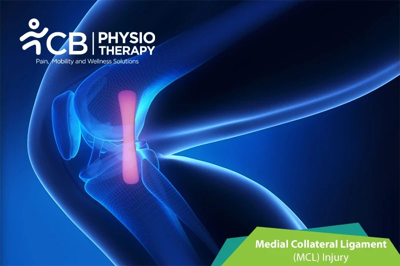
The MCL is located on the medial aspect of the knee and is attached to the thighbone and shinbone. The MCL holds the knee stable during valgus stress that is when stress is put on the outer part of the leg that can buckle the knee toward the center of the body.
Knee injuries that involve the MCL injuries are referred to as sprains or tears.
MCL sprains can be graded as follows:
MCL is usually related to the Medial meniscus and the anterior cruciate ligament (ACL), these two structures may also be damaged in association with an MCL injury.
Medial collateral ligament injuries are the most common ligament injuries of the knee. The reasons for the injury can be:
Symptoms vary from patient to patient depending on the severity of the injury. The symptoms can be:
Pathology
The medial collateral ligament is a disruption of the connection which is present between the medial femur and medial tibia. A medial collateral ligament sprain is graded as I, II, and III degrees. It may cause mild injuries with no laxity, or with some laxity, or with the ligaments in continuity, or those with complete disruption.
Physical examination:
Physical examination concentrates on the knee joint, hip joint, and ankle joint to identify any other associated injuries. Physical examination includes looking at the knee to check whether it is swollen and touching the knee in various places to check for pain and tenderness, the knee ligament is checked for its stability or joint laxity, by pushing on the outer side of the knee.
X-rays:
X-rays of the knee can be used to check fractures of the femur and tibia bones.
Stress X-ray:
The patient is kept in a relaxed position, and then the person doing the X-ray will gently pull on the MCL side of the knee to see if it opens up farther than it should. If the image shows a bigger gap between the shin and thigh bones than the normal, then the joint is loose, and the MCL is likely torn.
Magnetic resonance imaging (MRI):
Magnetic resonance imaging (MRI) is used to visualize the MCL and determine the grade of sprain, and how bad the strain or tear is.
Medication: Anti-inflammatory medications, pain killers, analgesics, etc.
Note: Medication should not be taken without the doctor's prescription.
Grade I and II sprain usually heal within one to two weeks. During the initial treatment, the patient is advised to wear a knee sleeve or hinged knee brace for comfort and protection, it also helps to increase range of motion and activity as tolerated by the patient. Patients will be usually recommended to use crutches for a few days.
Surgery: Usually grade III MCL sprain patients undergo surgery, but doctors only consider surgery in patients where conservative treatment has failed.
Rest:
Rest can be provided by preventing walking and other activities that cause pain.
Ice is applied for 15-20 minutes, every 2 hours a day. At about two or three weeks following injury, both the pain and swelling subside.
Compression:
The knee is kept in a knee brace or elastic bandage to compress the injured part.
Ultrasound therapy is used to reduce pain and increase the range of motion.
Transcutaneous electrical stimulation (TENS):
Transcutaneous electrical stimulations are used to decrease pain and swelling.
Elevation:
The needs to keep the affected knee elevated to keep the weight off the joint.
Crutches:
Crutches are used to assist while walking.
Range of motion exercises is used to help restore motion in the leg and the knee. After these exercises advanced stretching exercises are done, thus helping increase the range of motion.
Stretching exercises:
Stretching exercises like static stretching using a 30-second hold, relaxing, and performing 5 repetitions to hamstrings, iliotibial tract, and gastrocsoleus are recommended.
.
Strengthening exercises are recommended like quad sets, straight leg raises (to the front and to the back), hamstring curls, heel raises, heel dig bridging, shallow standing knee bends, etc. Isometric exercises progressed to isotonic and further isokinetic exercises are done gradually for strengthening.
Progressive resistive exercises (PRE):
Progressive resistive exercises are gradual self-resistive exercises with self-generated tension or graded resistance exercise with weight belts.
Balance exercises:
Balance exercises are used to enhance balance and equilibrium.
Plyometrics:
Plyometric exercise includes high-intensity training to cope with the high demands of sports activities. These exercises include jumping, running, reverse lunge knee-ups, squat jumps, clapping push-ups, etc.

Avoid twisting or pivoting the knee as it might be unstable. The patient should be careful while getting out of a car or catching the toe on a rug. Weight-bearing or walking should be started with the physiotherapist's instructions.
Get assessed today — find the cause, get a plan, and know when you'll feel better.