Verified
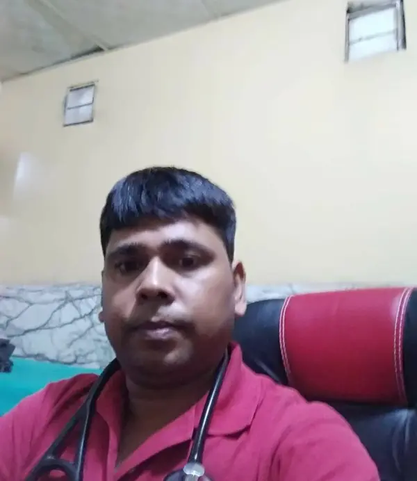
Dr. Kasim Saifi
Physiotherapist · BPT
See clinics, physiotherapists and prices for your city.
Whether it's a dull ache, a sharp catch, or pain that keeps coming back — we find the root cause of your Spina Bifida and fix it, at a clinic near you or at home.
Precision Care, Proven RecoveryWe'll call you within 10 minutes to confirm your home visit.We'll call you within 10 minutes to confirm your slot.
Chat with us on WhatsAppCommon triggers include muscle strain, joint stiffness, poor posture and long desk hours. If two or more of these fit, a physiotherapy assessment can identify exactly what's driving it.
Most spina bifida improves without surgery or long-term medication — the key is finding the cause first.
Spina Bifida rarely signals something serious, but see a doctor immediately if it comes with:
Our physios screen for these red flags at every first assessment and refer you to the right doctor if needed.
We start with an AI-based assessment to find the root cause — then follow a clear, four-phase plan.
Hands-on therapy and advanced modalities to calm pain and inflammation fast.
Mobility work and posture correction so everyday movement feels normal again.
Targeted strengthening that protects you — in-clinic or in our physio gym.
Back to work, sport and life — with a home program that prevents relapse.
One patient record, stage by stage: your physiotherapist pinpoints the diagnosis, finds the root cause and builds your in-clinic plan — while Fizo IQ™, our AI decision-support engine, tracks every milestone until your goals are met and you leave with an exercise plan to stay active.
These symptoms respond well to Spina Bifida — tap one to see how we treat it near you.
Make these exercises yours — a CB physio personalizes, doses and tracks your plan week by week.
Get my personalized planGeneral guidance only — stop if an exercise increases your pain, and check with your physio before starting a new routine.
Every CB physio is a qualified BPT/MPT clinician. You don't need to choose one — our lead physiotherapist reviews your case and assigns the specialist best suited to treat it.
Verified
Physiotherapist · BPT
Verified
Physiotherapist · BPT
Verified
Physiotherapist · BPT
Verified
Physiotherapist · MPT
Verified
Physiotherapist · BPT
Verified
Physiotherapist
Verified
Physiotherapist · BPT
Verified
Physiotherapist · BPT
Rated 5.0 / 5 across 31 verified patient reviews in Noida.
“Dr kasim listened well to my situation, was very quick to diagnose me and gave very clear guidelines on how I should manage my condition”
“I recently had the opportunity to experience physiotherapy at home, and I must say it was an outstanding and convenient experience. One of the biggest advantages of receiving physiotherapy at home was the convenience it offered. Instead of traveling to a clinic or hospital, the physiotherapists came to my doorstep, saving me time and energy. This personalized approach allowed me to receive treatment in the comfort of my own home, which greatly contributed to my overall well-being and recovery. Thanks Dr. kasim for the services.”
“I wholeheartedly recommend Dr. Kasim to anyone seeking treatment for a shoulder issue. His outstanding care, expertise, and compassionate approach make him an exceptional physiotherapist. Under his guidance, I experienced remarkable progress and successful recovery. Thank you, Dr. Kasim, for your exceptional care and dedication to helping your patients regain their shoulder health.”
“Dr. Vidhi's approach is highly professional and patient-centered. She took the time to thoroughly assess my condition, asking detailed questions and actively listening to my concerns. She ensured that I fully understood my diagnosis, the treatment plan, and the expected outcomes. . Her dedication to providing evidence-based care was evident. thanks for treated me so well..god bless.”
“Thank you sir for providing home physiotherapy treatment. its wonderful to have session with you. 🙏🙏”
“Hats off to you, ma'am. I am very satisfied with the treatment I had for body posture. Every session was great; it made me feel comfortable.👍👍”
Spina bifida is a neural defect that occurs during fetal development resulting from a defective fusion of one or more posterior vertebral arches causing protrusion of the contents of the spinal canal before birth, incomplete closing of the embryonic neural tube (a group of cells that form the brain and the spinal cord of a baby). This can cause physical and mental issues. The neural tube and spinal column do not develop properly and remain exposed along several vertebrae. The spinal cord and membranes are pushed out forming a sac on the back of the fetus. This sac may be covered with membranes or meninges.
Types of spina bifida:
Spina bifida occulta
There is merely a radiological defect between the laminae. Skin defects like haemangioma, lipoma, a tuft of hair, occur as a result of spina bifida occulta.
Spina bifida cystic
There is a developmental deficiency of laminae, spinous processes, overlying muscles, and skin.
Meningocele
In this condition mostly the spinal cord and its nerve roots are intact, only the covering membrane projects as a dural sac.
Myelomeningocele
It is associated with muscle paralysis, sensory loss, bladder and bowel incontinence, and deformities. It may be present from the 4th thoracic to 1st sacral vertebra. However, most common in the lumbosacral region. Associated with complications like paraplegia, deformities, bladder and kidney infections, skin ulceration, development of pathological fractures, meningitis as well as hydrocephalus.
Spina bifida can occur due to a combination of genetic, environmental, and nutritional factors.
The most severe symptoms of spina bifida are seen in the Myelomeningocele type of spina bifida. Other symptoms include:
Pathology
Incomplete development of the neural tube and spinal column and thus the spine does not close fully, and the spinal column remains exposed along several vertebrae. If the opening in the vertebrae occurs in the upper spine, there are chances of complete paralysis in the legs and others. If the openings are in the middle of the spine, symptoms tend to be less severe. There is fluid inside the brain and also around the surface of the brain and the spinal cord called cerebrospinal fluid (CSF). An increase in the CSF can lead to hydrocephalus, causing pressure on the brain, and brain damage.
Neural tube defects and other problems during pregnancy can be reduced by taking early action by these tests.
· Ultrasound
Ultrasound is used to scan the detects of spina bifida during pregnancy. Various prenatal tests can also be done, but they are not accurate.
· Alpha-fetoprotein (AFP) Blood test
This is a blood test that assesses for a protein, alpha-fetoprotein (AFP), that the fetus produces.
These tests are used to assess three or four substances in the blood.
· Amniocentesis
Test alpha-fetoprotein(AFP) levels are done by taking the sample of fluid from the amniotic sac. If there is a high level of AFP in the amniotic fluid then the fetus has a neural tube defect.
· Treatment
Treatment of spina bifida depends on its type and its severity.
· Surgical options
Surgery is possible in some cases.
· Surgery to repair the spine
The spinal cord and any exposed tissues or nerves are replaced and the gap is closed in the vertebrae the spinal cord with muscle sealed.
· Prenatal surgery
Usually, during 19TH to 25TH week of pregnancy. The spinal cord of the fetus is repaired by opening the uterus. This may reduce the risk of spina bifida after delivery.
· Hydrocephalus
A thin tube, or shunt, is implanted in the baby's brain are implanted. And the excess Cerebro Spinal fluid is drained into the abdomen.
· Medication
Anticholinergic, antibiotics, Botox injection.
· Physiotherapy treatment includes
· Prevention and management of deformities:
Less severe types of deformities may be treated by passive stretching and adequate splinting. Rigid deformities may need manipulation under general anesthesia, followed by plaster and splints.
· Physiotherapy treatment after surgery:
Reeducate the transplanted muscle iliopsoas to function as an effective hip abductor and extensor is of primary importance. ROM range of motion exercises and muscle strengthening at the knee to offer stabilization in standing and walking.
· Management of muscle paralysis:
Muscles like hip abductors, extensors, knee extensors, ankle dorsi flexors, and plantar flexors should be strengthened for weight-bearing. Also strengthen the shoulder girdle and trunk muscles, as they also are functionally important.
Prevent adverse factors like hip flexion, knee flexion, and ankle dorsiflexion contractures by emphasizing hip extension, knee extension, and ankle plantigrade programs.
· Balance and coordination exercises:
To maintain body equilibrium in various positions, arm muscles, visual and auditory feedback techniques can be used. Reeducation of the trunk muscles is required in children with trunk lesions.
· Care of skin and joints:
Proper checking of the splint and brace by frequent careful examination of the skin at pressure points. Parents should also be careful about exposing the insensitive parts of the limb to extreme hot and cold situations.
· Education in ambulation and self-care:
Develop equilibrium, static and dynamic weight-bearing programs. Re-educate in standing and gait training.
Arm and shoulder muscles are strengthened for crutch walking.
· Assistive technologies:
An electric wheelchair is provided to the person with total paralysis of the legs.
· Special leg braces:
Leg braces help to prevent the lower limb muscles from weakening and make the patient independent.

Get assessed today — find the cause, get a plan, and know when you'll feel better.