Open today · 8 AM–8 PM · Same-day slots
CB Physiotherapy
Same-day appointments

Quadriceps Muscle Strain Treatment Doctors in Jammu And Kashmir

Whether it's a dull ache, a sharp catch, or pain that keeps coming back — we find the root cause of your Quadriceps Muscle Strain and fix it, at a clinic near you or at home.

Precision Care, Proven Recovery
ACCURATE DIAGNOSIS
ROOT CAUSE ASSESSMENT
COMPREHENSIVE RECOVERY PLAN
MILESTONE TRACKING
GOALS ACHIEVED
STAYING ACTIVE
See how it works ↓
Move pain-free Avoid drugs & surgery
5.0 / 5 · 23 Google reviews Verified on Google

Request received!

We'll call you within 10 minutes to confirm your home visit.We'll call you within 10 minutes to confirm your slot.

Chat with us on WhatsApp

Book in 30 seconds

Visit your nearest CB clinic. Tell us your concern and a preferred time — we'll confirm your slot by call within 10 minutes.

Same-day booking by default. We'll confirm your exact slot by call.
+91
No spam · Your number is safe with us
Prefer to talk now? Call +08082666216
Understanding your quadriceps muscle strain

Does this sound like you?

Common triggers include muscle strain, joint stiffness, poor posture and long desk hours. If two or more of these fit, a physiotherapy assessment can identify exactly what's driving it.

  • A dull ache or sharp, burning quadriceps muscle strain that keeps returning
  • Discomfort that worsens with movement, lifting or long sitting hours
  • Morning stiffness that eases as you move
  • Pain that limits work, sport or everyday activities
  • Recurring episodes that keep coming back

Most quadriceps muscle strain improves without surgery or long-term medication — the key is finding the cause first.

⚠ When to seek urgent medical care

Quadriceps Muscle Strain rarely signals something serious, but see a doctor immediately if it comes with:

  • Fever, unexplained weight loss, or a history of cancer
  • Numbness or weakness that keeps getting worse
  • Loss of bladder or bowel control
  • Pain after a major fall or accident

Our physios screen for these red flags at every first assessment and refer you to the right doctor if needed.

Assessment-led treatment

How we treat quadriceps muscle strain

We start with an AI-based assessment to find the root cause — then follow a clear, four-phase plan.

Phase 1

Ease the pain

Hands-on therapy and advanced modalities to calm pain and inflammation fast.

Phase 2

Restore movement

Mobility work and posture correction so everyday movement feels normal again.

Phase 3

Build strength

Targeted strengthening that protects you — in-clinic or in our physio gym.

Phase 4

Return to activity

Back to work, sport and life — with a home program that prevents relapse.

Your physiotherapist, with Fizo IQ™

Every recovery, planned and proven

One patient record, stage by stage: your physiotherapist pinpoints the diagnosis, finds the root cause and builds your in-clinic plan — while Fizo IQ™, our AI decision-support engine, tracks every milestone until your goals are met and you leave with an exercise plan to stay active.

Physio Expert @CB with Fizo IQ™ — recovery engine
TRACKING
ACCURATE DIAGNOSIS
L4–L5 disc bulge · nerve-root irritation, pinpointed at the first movement assessment
ROOT CAUSE ASSESSMENT
Poor sitting posture, weak core–hip chain · quietly loading that disc
COMPREHENSIVE RECOVERY PLAN
24 clinic sessions · manual therapy + spinal decompression · guided exercise therapy, reviewed weekly
MILESTONE TRACKING
Sitting pain-free ✓ · stairs ✓ · next: the morning 5 km walk
GOALS ACHIEVED
Pain 7 → 3 in six weeks · back to daily life, moving freely
STAYING ACTIVE
A personalized exercise plan, yours to keep · so the pain doesn't come back
Example recovery journey · every plan is built around you
Move better

Exercises that help with Quadriceps Muscle Strain

More exercises →

Make these exercises yours

Get my personalized plan

General guidance only — stop if an exercise increases your pain, and check with your physio before starting a new routine.

Meet your care team

Licensed experts who treat hundreds like you every month

Every CB physio is a qualified BPT/MPT clinician. You don't need to choose one — our lead physiotherapist reviews your case and assigns the specialist best suited to treat it.

Dr. Shafiya, physiotherapist in Anantnag

Dr. Shafiya

Physiotherapist

5.0 (13)
Anantnag
cb physiotherapy janglat mandi
1 yrs experience
Ortho Spine Sports Physiotherapy Center
Dr. Athur Un Nisa, physiotherapist in Anantnag Verified

Dr. Athur Un Nisa

Physiotherapist

5.0 (10)
Anantnag
cb physiotherapy janglat mandi
4 yrs experience
Ortho Spine Sports Physiotherapy Center
Not sure who to see? You don't have to decide. Our lead physiotherapist reviews every case and assigns the physio best suited to it — based on your condition and availability.
Patient stories

Quadriceps Muscle Strain recoveries, verified on Google

Rated 5.0 / 5 across 23 verified patient reviews in Jammu And Kashmir.

Verified

“Received treatment at Cb Anantnag clinic where Dr. Athar treated me she was highly skilled and professional. Her treatment approach is effective, and I noticed significant improvement in my knee pain within a short time. I highly recommend her for quality physiotherapy care. in Anantnag”

KZ
Khan Zainab Verified patient · Treated at CB · 7 months ago
Verified

“Excellent physiotherapist, Dr. Athar! Her expertise and customized care assisted me in recovering swiftly. from back issue, I strongly recommend her..”

SJ
Saima Jan Verified patient · Treated at CB · 7 months ago
Verified

“I had an excellent experience with Dr. Shafiya. My back pain recovery was made easier and less stressful thanks to her knowledge and kind attitude. Thank you for your incredible care. Doctor Shafiya.”

SB
Saima Bhat Verified patient · Treated at CB · 7 months ago
Verified

“Dr. Shafiya is an excellent physiotherapist! Her expertise, concern, and dedication enabled me to heal fast and painlessly. I definitely recommend her to anyone looking for efficient physiotherapy treatment.”

RJ
Rumaisa Jaan Verified patient · Treated at CB · 7 months ago
Verified

“Dr. Athar un Nisa is a wonderful physical therapist who honestly cares about her patients. She devised a treatment regimen that helped me restore mobility and strength following my injury. Her treatment sessions were quite successful and motivational. I am incredibly appreciative for her effort and support.”

RB
Rafia Bhat Verified patient · Treated at CB · 7 months ago
Verified

“Dr. Athar un Nisa is a wonderful physical therapist who honestly cares about her patients. She devised a treatment regimen that helped me restore mobility and strength following my injury. Her treatment sessions were quite successful and motivational. I am incredibly appreciative for her effort and support.”

RB
Rafia Bhat Verified patient · Treated at CB · 7 months ago
Good to know

Quadriceps Muscle Strain, answered

A quadriceps muscle strain is an acute tearing injury of the quadriceps, which usually occurs due to an acute stretch of the muscle and forceful contraction at the same time or repetitive functional overloading.

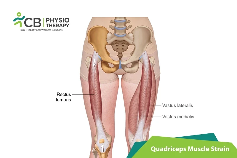
The Quadriceps femoris muscle is located in the anterior compartment of the thigh and is a hip flexor and a knee extensor. It consists of four parts:

  • Rectus femoris
  • Vastus lateralis
  • Vastus medialis
  • Vastus intermedius

Grades of Quadriceps Strain:

Grade 1: Grade 1 quadriceps strain is not always a serious injury. Spasm is at the site of injury and feeling of tightness and mild discomfort on walking and running might be difficult.

Grade 2: The patient feels a sudden sharp pain while running, jumping, or kicking and is unable to play. Difficulty in walking with swelling or mild bruising is present. Pain is felt while pressing in on the location of the quad muscle tear. Straightening of the knee against resistance causes pain and the patient is unable to fully bend the knee.

Grade 3: The patient feels severe, sudden pain in the front of the thigh and is unable to walk without a walking aid. Severe swelling appears immediately with bruising within 24 hours. Muscle contraction will be painful and can produce a bulge in the muscle.

Among quadriceps muscles, the rectus femoris is most frequently strained, due to reasons such as muscles crossing two joints, those with a high percentage of Type II fibers, and muscles with complex musculotendinous architecture. Acute injuries usually occur as a result of:

  • Sudden deceleration movement of the leg,
  • Violent contraction of the quadriceps,
  • Rapid deceleration of an overstretched muscle while quickly changing the direction,
  • Muscle fatigue,
  • Traumatic event, macro-trauma to the muscle,
  • Due to contact sports such as rugby, soccer, and basketball, etc
  • Overloaded by repeated eccentric muscle contractions of the knee extensor mechanics,
  • As a result of overuse,
  • Repetitive micro-trauma to the muscle,
  • Excessive passive stretching can also cause strains.

There is an increased risk of rupture due to a few medical conditions or due to the use of certain medications, including the following:

  • Rheumatoid arthritis,
  • Kidney disease,
  • Diabetes mellitus
  • Metabolic bone disease (such as hyperparathyroidism),
  • Gout,
  • Obesity,
  • Systemic lupus erythematosus (SLE), etc.

Symptoms of quadriceps strain depend on the type of injury, though common symptoms can be:

  • Pain while stretching, flexing, or using the quads for movement,
  • Swelling,
  • Muscle spasm,
  • Bruising as a result of broken blood vessels in the injured area,
  • Loss of strength,
  • Cracking sensation while pushing the fingers on the injured area of the thigh.

Pathology:

Strains are seen in all the quadriceps muscles but the rectus femoris is most vulnerable to strain as it passes over two joints: the hip and the knee. The most common site of strain is the distal musculotendinous junction of the rectus femoris. The patient feels a sudden pain in the anterior thigh during an activity requiring excessive muscle contraction. Other symptoms are local pain, tenderness, severe swelling, and bruising.

Physical examination:

After obtaining a thorough medical history, careful physical examination is done for observation, palpation, strength testing, and evaluation of motion.

The examiner has a close look at the affected area, observes swelling and bruising. Also, observes the patient in standing and walking, looks for postural abnormalities. Palpation of the quadriceps muscle is done along the entire length of the muscles. It's required to identify swelling, thickening, tenderness, defects, and masses if present. Strength testing of the quadriceps includes the resistance of knee extension and hip flexion. Adequate strength testing of the rectus femoris includes resisted knee extension with the hip flexed and extended. For this, the best way to evaluate the patient is in both sitting and prone-lying position. The prone-lying position allows optimum assessment of quadriceps motion and flexibility. Pain is usually felt by the patient with resisted muscle activation, passive stretching, and direct palpation over the muscle strain.

Ely's test:

The Ely's test is a physical test used to assess rectus femoris spasticity or tightness. The patient lies prone and the therapist stands next to the patient, at the side of the leg that is to be tested. One hand is placed on the lower back, the other hand holds the leg at the heel. Then flex the knee passively, the heel should touch the buttocks. For comparison test both sides. The test is positive if the heel cannot touch the buttocks, and the hip of the tested side rises from the table, the patient feels pain or tingling in the back or legs.

Radiographs:

Radiographs are effective to differentiate the etiology of the pain in the quadriceps muscles, which can be a muscular strain or stress fracture etc.

Ultrasound:

Ultrasound is often used as it is inexpensive. But it also has a disadvantage, that it's highly operator dependent and requires a skilled and experienced clinician. An advantage is that it can image the muscles dynamically and assess for bleedings and hematoma formation via Doppler.

Magnetic resonance imaging (MRI):

Magnetic resonance imaging (MRI) is used to give detailed images of muscle injury.

Medication: NSAIDs, pain killers, analgesics, etc.

Note: Medication should not be taken without the doctor's prescription.

Surgery:

Conservative treatment results in a good outcome. But there are certain specific indications in which surgical intervention might be beneficial, these are:

  • Large intramuscular hematoma,
  • Complete (III degree) strain,
  • Tear of a muscle,
  • Partial or II degree strain if more than half of the muscle belly is torn.

Rest:

Rest prevents worsening of the injury. Place the injured knee to rest the first few days after the trauma, this can prevent the formation of a large gap within the muscle, reduce the size of the hematoma, and the size of the connective tissue scar. A short period of immobilization accelerates the formation of granulation tissue at the site of injury during the initial phase. Gradually mobilization should be started to optimize the healing by restoring the strength of the injured muscle, preventing muscle atrophy, the loss of strength, and extensibility.

Ice therapy:

Ice therapy lowers the intra-muscular temperature and decreases blood flow.

Compression:

Compression wrap or bandage can help to decrease both blood flow and excess interstitial fluid accumulation. It aims to prevent hematoma formation and interstitial edema, thus decreases tissue ischemia.

Elevation:

The elevation of an affected leg above the level of the heart results in a decrease in hydrostatic pressure, and also reduces the accumulation of interstitial fluid, so there is less swelling at the place of injury.

Therapeutic Ultrasound:

Ultrasound helps to enhance the process of healing. It produces the thermal effect, which is of benefit to chronic muscle strains, it also reduces inflammation and promotes healing, which can help with an acute injury.

Transcutaneous electrical stimulation (TENS):

Transcutaneous electrical stimulation (TENS) can be used to decrease pain and swelling. It can help pump out inflammation, reduce pain, and desensitize nerve fibers. It also can strengthen muscle and re-educate.

Shockwave therapy:

Shockwave therapy is used to produce an improvement in healing and regeneration by a single acoustic wave impulse which stimulates healing of soft tissue, mainly by inhibiting the function of afferent pain receptors and increasing angiogenesis. Therefore, it increases blood flow by inducing a healing process mediated by the generated inflammatory process

Mobilization:

Mobilization is done to restore normal pain-free movement of the leg. The areas above and below the injury site are mobilized initially. It should be carried out gently and initially do not mobilize over the site of injury. But as the pain decreases, mobilization over adjacent sites to the injury can be started gently.

Active range of motion exercises:

Active range of motion is done once the injured leg is recovered well. This phase includes simple range of motion exercises like knee flexion extension, ankle movement, and toe movements. But before starting with AROM exercises passive movements are recommended.

Stretching Exercises:

Stretching exercises should be done carefully to the point of discomfort, but without pain. This includes techniques such as passive, active-passive, dynamic, and proprioceptive neuromuscular facilitation stretching. e.g. Static quad stretch, hip flexor stretch, etc.

Strengthening Exercises:

Strengthening exercises are done to gradually increase the load that is put through a muscle. Initially, isometric or static exercises are advised, then progress to low-level pain-free exercises and then to advanced exercises with the resistance band and finishing with sports-specific running and sprint drills. Strengthening exercises can also be done in an aquatic pool by using the resistance of water.

Isotonic Exercise:

Isotonic exercises are begun with the lightest free weight, 3 sets of 10 repetitions up to 3 times a day. Terminal knee extensions at 20 degrees, increase as comfort and knee flexion allows. Once terminal knee extensions are done properly without extensor lag, free weights are added to the SLRs and terminal knee extensions, weight is increased slowly.

Eccentric Exercises:

These exercises include higher velocity eccentric exercises that include plyometric and sports specific activities such as squat jumps, split jumps, bounding and depth jumps, single leg bounding, backward skips, lateral hops, lateral bounding, plyometric box jumps, eccentric forward pulls, eccentric backward steps, eccentric lunge drops, single and double leg deadlifts, etc.

Isokinetic Exercises:

Isokinetic exercises are very high-intensity exercises such as swimming, running, treadmill walking Isokinetic strength testing should be performed under both concentric and eccentric action conditions.

The patient should be advised to do a proper warm-up and stretching before exercises or post-exercise. There should be a gradual increase in the exercise intensity or volume with complete monitoring. Complete rehabilitation programs should be followed in case of any injury to the lower limb.

Ready when you are

Don't let the pain decide your day.

Get assessed today — find the cause, get a plan, and know when you'll feel better.

📞 Call 💬 WhatsApp Book now