
Lateral Collateral Ligament(lcl) Injury — Symptoms, Causes & Treatment
Also known as Lateral Collateral Ligament(lcl) Injury. Causes, symptoms and how physiotherapy treats Lateral Collateral Ligament(lcl) Injury — without surgery or medicines.
Understanding Lateral Collateral Ligament(lcl) Injury
Lateral Collateral Ligament(lcl) Injury is one of the conditions our physiotherapists treat most often — and one of the most treatable when the actual driver is found. Two people with Lateral Collateral Ligament(lcl) Injury rarely share the same underlying cause, which is why one-size-fits-all treatment so often disappoints.
What matters clinically is the pattern: how it started, what makes it better or worse, and how it behaves through the day. That pattern — mapped in a structured assessment — tells us which structures are involved and which treatment will actually move the needle.
Causes & risk factors of Lateral Collateral Ligament(lcl) Injury
What commonly drives it — your assessment pins down which of these apply to you.
Sudden spikes in activity
A new sport, a new gym program, a long trek — tissue adapts to gradual change and protests when change is sudden.
Muscle strain and overload
A sudden lift, an awkward movement or simply more load than the tissues were prepared for — the most common trigger.
Posture and long static hours
Desks, driving and screens keep the same structures loaded for hours at a stretch — comfortable at first, aching later.
Joint stiffness and age-related change
Joints that move less start to hurt more; age-related wear is normal, but painful stiffness is not something to simply accept.
Weakness and deconditioning
When supporting muscles lose strength, load shifts to structures that were never meant to carry it.
When physiotherapy is the right call
If any of these sound like you, an assessment is the right first step.
- ✓ Pain or stiffness that limits work, sleep or daily activities
- ✓ You want to avoid long-term painkillers — or surgery has been mentioned
- ✓ You've been diagnosed with Lateral Collateral Ligament(lcl) Injury and told to "rest and see"
- ✓ Movement feels restricted, weak or guarded compared to before
- ✓ You want to return to sport or work with a plan, not by trial and error
Your treatment journey
Step by step — from your first assessment to a tracked recovery plan.
A detailed first assessment
History, movement testing and strength checks — a focused session to find what is actually driving your Lateral Collateral Ligament(lcl) Injury.
A diagnosis you understand
Your physiotherapist explains the findings in plain language — what is affected, why it happened and what that means for you.
Active, tracked treatment
Hands-on therapy, the right modalities and graded exercise — progressed session by session and tracked on Fizo IQ™.
Prevention, built in
The final phase rebuilds strength and habits so the same problem does not come back — with a home plan you keep.
What recovery looks like
- ✓ Relief that comes from treating the cause, not masking it
- ✓ A clear diagnosis and a timeline you can plan around
- ✓ Strength and movement rebuilt, not just pain reduced
- ✓ Lower chance of recurrence, with a prevention plan
- ✓ Care without long-term medicines — at a clinic near you or at home
When to see a doctor — red flags
How CB Physiotherapy treats Lateral Collateral Ligament(lcl) Injury
We treat Lateral Collateral Ligament(lcl) Injury the way we treat everything: assessment first, plan second, treatment third. It sounds obvious, but it is the difference between sessions that fill a slot and sessions that move you towards a goal. Care is delivered by certified physiotherapists at a clinic near you or at home.
Your whole journey is tracked on Fizo IQ™ — the diagnosis, the root cause, the milestones and the goals — so progress is measured, not guessed, and the plan adapts the moment your response calls for it.
Exercises that help with Lateral Collateral Ligament(lcl) Injury
Make these exercises yours — a CB physio personalizes, doses and tracks your plan week by week.
Get my personalized planGeneral guidance only — stop if an exercise increases your pain, and check with your physio before starting a new routine.
Get Lateral Collateral Ligament(lcl) Injury treatment near you
Certified physiotherapists at a clinic near you or at home — same-day slots, assessment-led care.
Request received!
We'll call you within 10 minutes to confirm your home visit.We'll call you within 10 minutes to confirm your slot.
Chat with us on WhatsAppLateral Collateral Ligament(lcl) Injury, answered
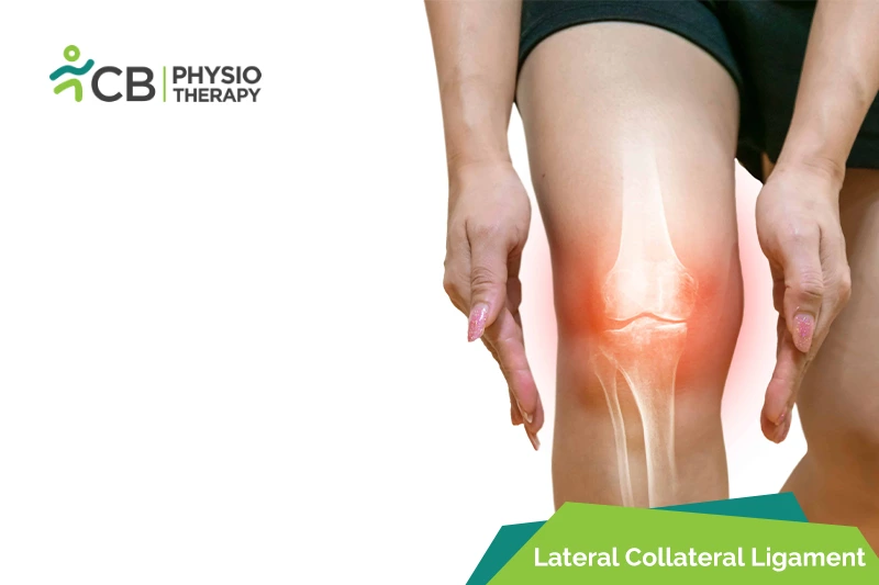
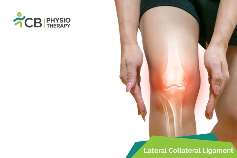
Knee joint is the largest joint in the body and is one of the easiest joints to get injured. Lateral collateral ligament (LCL) sprains and tears, however, are much less common knee injuries. The knee's collateral ligaments are the lateral collateral ligament (LCL) and the medial collateral ligament (MCL), located on either side of the knee. These ligaments control the sideways motion of the joint, protecting it from unusual movement side-to-side. The lateral collateral ligament (LCL) runs along the outer side of the knee.
Types of lateral collateral ligament (LCL) sprain:
Grade 1 lateral collateral ligament (LCL) sprain, the ligament is stretched, with mild to moderate pain, mildly injured, and minimally decreased range of motion. But is still able to keep the knee stable and functional.
Grade 2 Lateral collateral ligament (LCL) sprain or partial LCL tear, with severe pain, the ligament is stretched or stressed and becomes loose and range of motion is moderate to severe decreased.
Grade 3 Lateral collateral ligament (LCL) sprain or complete LCL tear with severe pain, the ligament is torn into two separate pieces and the joint is no longer stable and the range of motion is severely decreased.
The knee joint is vulnerable to hard muscle contraction and direct impact, caused by:
- Quick cuts,
- Sudden changing direction while running,
- The movement that causes inward stress on the knee,
- Direct contact or force, jumping, twisting, and quick changes in direction,
- Lateral movements of the knee to unnatural motion,
- Occurs mostly in soccer players, football players, basketball players, and skiers.
Lateral collateral ligament (LCL) sprains symptoms can vary in severity, depending on the grade of injury. Symptoms include:
- Mild or acute pain,
- Swelling on the lateral side of the knee,
- Tenderness or bruising on the outer side of the knee,
- Discoloration/bruising,
- Decreased range of motion,
- Instability,
- Locking in the joint when moved,
- Joint stiffness,
- Feeling of popping" or snapping,
- Feeling of instability or giving out underweight.
Pathology:
The lateral collateral ligament injury is a disruption of the connection which is present between the lateral femur and lateral fibula. A lateral collateral ligament sprain is graded as I, II, and III degrees. It may cause mild injuries with some or no laxity, with the ligaments in continuity, or with complete disruption.
Physical examination:
Physical examination includes observing the swelling, ecchymosis, and warmth along the lateral joint line. The patient is assessed for range of motion, muscle strength, sensation, reflexes, and palpation along the lateral joint line. Gait analysis and special tests are performed to determine associated ligamentous, meniscal, or soft tissue injuries.
X-ray:
X-ray of AP and Lateral side is used to rule out associated structural injuries. Varus and Posterior kneeling stress images are used to determine injury of LCL.
Magnetic resonance imaging (MRI):
Magnetic resonance imaging (MRI) is the gold standard in diagnosing LCL injuries. Coronal and Sagittal images are used in diagnosing an LCL injury.
Ultrasound is used in case of rapid diagnosis of LCL injury. Thickened and hypoechoic LCL indicates an LCL injury. In case of a complete tear, an ultrasound may show increased edema, lack of fiber continuity, dynamic laxity of the LCL.
Medication: Nonsteroidal anti-inflammatory drugs (NSAIDs), pain killers, analgesics, etc.
Note: Medication should not be taken without the doctor's prescription.
Surgery:
Lateral Collateral ligament (LCL) injuries tend to involve other structures of the knee that might require additional treatment. Some LCL sprains might heal by conservative treatment others require surgery.
Surgery is performed as an open procedure combined with arthroscopy. Torn LCL is replaced using a tissue graft, which is passed through tunnels within the bone and then attached to the fibula and femur bones using screws.
Reconstruction is another technique used, it involves an allograft hamstring tendon or an autograph to reconstruct the LCL. Usually, reconstruction of the LCL in the knee with a patellar tendon allograft is also done.
Rest:
Rest is recommended to prevent the injured area from any stress and to protect it from further injury. This knee brace, may be helpful to protect the ligament from further sideways movements can also help speed the healing process.
Ice applied immediately after injury to the injured area for 15 to 20 minutes at a time every hour can help reduce swelling and inflammation of the joint.
Compression:
After cryotherapy, a compression sleeve is worn, this helps to control swelling and protects the knee.
Elevation:
Elevation promotes circulation of the blood back to the heart, this helps to drain the toxic substances as the injured tissue is broken down by the body.
Ultrasound therapy is used to reduce pain and increase the range of motion.
Transcutaneous electrical stimulation (TENS):
Transcutaneous electrical stimulations (TENS) are used to decrease pain and swelling.
Range of motion exercises:
Range of motion exercises are started in pain-free ranges of motion. Physiotherapist teaches specific exercises to help restore function while improving strength and stability. E.g lie on the stomach with both legs straight, bend one knee up as far as possible, and then straighten out so it is straight again. Repeat this exercise 10-20 times in a pain-free range.
Physiotherapy helps to strengthen the muscles around the knee. E.g. take a resistance band and put it on the lower leg, feet should be kept hip-width apart, then squat down and step out with one leg wider than the hip-width, then bring afterward the other leg, return to hip-width. Repeat this exercise 10 times in each direction and should be done in a pain-free range of motion.
Stretching exercises:
Stretching exercises are used to increase joint range of motion. E.g. bring the foot up towards the buttock with the same side hand, then push the hips forward and leg back, and feel stretch on the front of the thigh and hold it for 30 seconds repeat 3 times.
Progressive resistive exercises (PRE):
Progressive resistive exercises are recommended to the patient, these are gradual self-resistive exercises with graded resistance with weight belts or self-generated tension.
Balance exercises:
Balance exercises are done with the help of a wobble board which is used to enhance balance and equilibrium.
Plyometrics:
Plyometric exercises are high-intensity exercises used to cope with high demands like sports activities. Examples are jumping, running, reverse lunge knee-ups, squat jumps, clapping push-ups, etc.
The patient is advised to continue doing the strengthening exercises to prevent the knee from re-injury. The patient can continue to wear a brace while activities.
Don't let Lateral Collateral Ligament(lcl) Injury decide your day.
Get assessed today — find the cause, get a plan, and know when you'll feel better.