Open today · 8 AM–8 PM · Same-day slots
CB Physiotherapy
Same-day appointments

Flat Foot Treatment Doctors in Hyderabad

Whether it's a dull ache, a sharp catch, or pain that keeps coming back — we find the root cause of your Flat Foot and fix it, at a clinic near you or at home.

Precision Care, Proven Recovery
ACCURATE DIAGNOSIS
ROOT CAUSE ASSESSMENT
COMPREHENSIVE RECOVERY PLAN
MILESTONE TRACKING
GOALS ACHIEVED
STAYING ACTIVE
See how it works ↓
Move pain-free Avoid drugs & surgery

Request received!

We'll call you within 10 minutes to confirm your home visit.We'll call you within 10 minutes to confirm your slot.

Chat with us on WhatsApp

Book in 30 seconds

Visit your nearest CB clinic. Tell us your concern and a preferred time — we'll confirm your slot by call within 10 minutes.

Same-day booking by default. We'll confirm your exact slot by call.
+91
No spam · Your number is safe with us
Prefer to talk now? Call +04045210121
Understanding your flat foot

Does this sound like you?

Common triggers include muscle strain, joint stiffness, poor posture and long desk hours. If two or more of these fit, a physiotherapy assessment can identify exactly what's driving it.

  • A dull ache or sharp, burning flat foot that keeps returning
  • Discomfort that worsens with movement, lifting or long sitting hours
  • Morning stiffness that eases as you move
  • Pain that limits work, sport or everyday activities
  • Recurring episodes that keep coming back

Most flat foot improves without surgery or long-term medication — the key is finding the cause first.

⚠ When to seek urgent medical care

Flat Foot rarely signals something serious, but see a doctor immediately if it comes with:

  • Fever, unexplained weight loss, or a history of cancer
  • Numbness or weakness that keeps getting worse
  • Loss of bladder or bowel control
  • Pain after a major fall or accident

Our physios screen for these red flags at every first assessment and refer you to the right doctor if needed.

Assessment-led treatment

How we treat flat foot

We start with an AI-based assessment to find the root cause — then follow a clear, four-phase plan.

Phase 1

Ease the pain

Hands-on therapy and advanced modalities to calm pain and inflammation fast.

Phase 2

Restore movement

Mobility work and posture correction so everyday movement feels normal again.

Phase 3

Build strength

Targeted strengthening that protects you — in-clinic or in our physio gym.

Phase 4

Return to activity

Back to work, sport and life — with a home program that prevents relapse.

Your physiotherapist, with Fizo IQ™

Every recovery, planned and proven

One patient record, stage by stage: your physiotherapist pinpoints the diagnosis, finds the root cause and builds your in-clinic plan — while Fizo IQ™, our AI decision-support engine, tracks every milestone until your goals are met and you leave with an exercise plan to stay active.

Physio Expert @CB with Fizo IQ™ — recovery engine
TRACKING
ACCURATE DIAGNOSIS
L4–L5 disc bulge · nerve-root irritation, pinpointed at the first movement assessment
ROOT CAUSE ASSESSMENT
Poor sitting posture, weak core–hip chain · quietly loading that disc
COMPREHENSIVE RECOVERY PLAN
24 clinic sessions · manual therapy + spinal decompression · guided exercise therapy, reviewed weekly
MILESTONE TRACKING
Sitting pain-free ✓ · stairs ✓ · next: the morning 5 km walk
GOALS ACHIEVED
Pain 7 → 3 in six weeks · back to daily life, moving freely
STAYING ACTIVE
A personalized exercise plan, yours to keep · so the pain doesn't come back
Example recovery journey · every plan is built around you
Move better

Exercises that help with Flat Foot

More exercises →

Make these exercises yours

Get my personalized plan

General guidance only — stop if an exercise increases your pain, and check with your physio before starting a new routine.

Meet your care team

Licensed experts who treat hundreds like you every month

Every CB physio is a qualified BPT/MPT clinician. You don't need to choose one — our lead physiotherapist reviews your case and assigns the specialist best suited to treat it.

Not sure who to see? You don't have to decide. Our lead physiotherapist reviews every case and assigns the physio best suited to it — based on your condition and availability.
Good to know

Flat Foot, answered

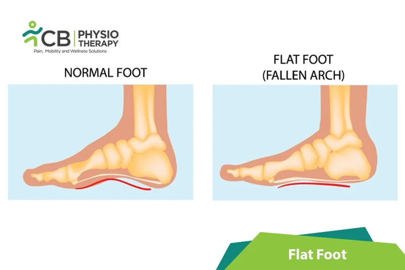
A flat foot is a deformity that occurs when the arch of the foot collapses and comes into contact with the ground. It is also known as pes planus deformity and may occur at the time of birth or may develop over time, mostly due to age or injury. Foot arches add elasticity and flexibility and are also very important for proper foot movement. A fallen arch may cause intense foot pain in the feet, ankles, and lower leg muscles.

Types of the Flat Foot:

Flexible Flat Feet (FFF) and Rigid Flat Feet (RFF).

Flexible Flat Feet (FFF) or pediatric flat feet are first apparent in childhood. It occurs due to weak ligaments that connect the bones of the foot, or due to muscle or tendon weakness. It is usually present in both feet. The arch is absent in the standing position and re-appears when the foot is in any other position.

Rigid Flat Feet (RFF) or adult acquired flat feet occur in adults. It develops due to the weakening of the tibialis posterior muscle tendon and leads to flattening of the arch and rolling of the ankle. It is a progressive deformity and causes pain and swelling on the inside of the foot.

Most people with flat foot go unnoticed as this condition may not cause any symptoms. If present, the most common symptoms of the flat foot include:

  • Pain around the areas of the arches, heels, and ankles,
  • The pain usually increases with activity,
  • Swelling occurs along the inner ankle and arch,
  • Hip, knee, and lower back pain present as instability of gait and posture can place undue stress on these joints,
  • Certain foot movements like standing on toes can become unbearable,
  • There can be a limitation in the range of motion between the bones of the foot while walking or running, which can lead to a flat-footed gait.

Pathology:

A flat foot occurs due to the weakness of the muscular, ligamentous, or bony arch-supporting structures which leads to the collapse of the arch. Factors that contribute to an acquired flat foot deformity are obesity excessive tension in the triceps surae, posterior tibial tendon dysfunction, or ligamentous laxity in the spring ligament, plantar fascia, or other supporting plantar ligaments.

Flat feet may occur due to wear and tear of the tissues (ligaments and tendons) holding the joints in the foot. Whereas in some children, the arches may fail to develop due to various reasons. The common causes of the flat foot include:

  • Genetic disorders like calcaneovalgus, congenitally fused foot bones, etc,
  • Nerve problems or muscle diseases such as muscular dystrophy, cerebral palsy, or spina bifida,
  • Aging,
  • Limb length inequality,
  • Standing or walking for long periods,
  • Weakened muscles in the foot, ankles, and lower leg,
  • Pregnancy,
  • Past injuries like a stress fracture,
  • Marfan's syndrome, Rheumatoid arthritis, scoliosis,
  • Excessive pronation of the foot,
  • Deterioration of the posterior tibial tendon, which runs along the inner ankle,
  • Ligamentous laxity,
  • Hypermobility,
  • Metatarsus adductus,
  • Hypertension and diabetes,
  • Obesity,
  • Inappropriate shoes like compressed toe box.

Physical examination:

The examiner evaluates the health history of the patient for evidence of any illnesses or foot problems that may be linked to flat foot. The feet are observed while toe standing, which might indicate flexible flatfoot, or the appearance of pain while standing or walking. The soles of the shoes are checked for any unusual patterns.

Wet footprint test:

This test is performed by wetting the feet and standing on a smooth, level surface. The thicker the print between the ball of the foot and the heel, the flatter the foot. Normally a high arched foot leaves only a narrow print of the outer foot.

Shoe inspection test:

This test is done to check the faulty foot mechanics. In case of flat foot, there will be more wear on the inside of the sole, especially in the heel area. Shoe's upper part will also tend to lean inward over the sole.

Too many toes test:

This test is performed as the examiner stands behind the patient and counts the number of toes peeking out to the sides. While only the little toe would be seen in people with normal pronation, 3 or 4 may be seen in those who overpronate.

Tiptoe test:

This test is used to see if the patient has flexible or rigid flat feet. If a visible arch is formed when the patient stands on the toes, then there is a flexible flat foot. If not, then the patient has a rigid flat foot.

X-rays and computed tomography (CT) scans:

These tests are used for diagnosing arthritis and evaluating irregularities in the angle or alignment of the foot bones.

Ultrasound:

Ultrasound is used to produce detailed images of soft tissue damage, such as a ruptured tendon.

Magnetic resonance imaging (MRI):

MRI helps to provide detailed images of bone and soft tissue damage, ideal for people with tendonitis, rheumatoid arthritis, or an Achilles heel injury.

Medication: Non-steroidal anti-inflammatory medications (NSAIDs), corticosteroids, etc.

Note: Medication should not be taken without the doctor's prescription.

Treatment of flat foot differs based on the person's age and cause. Flat foot in children will often resolve without intervention, while flat foot in adults tend to be permanent and non-reversible. In newborns and toddlers, a flat foot is normal because the arches haven't yet developed. While most children will develop arches by the age of 3, some may have structural deformities or experience delays that interfere with the normal alignment of the foot bones and may come and go as a child develops.

Whereas in adults flat foot is almost always permanent. People with a flexible flat foot in which the arch is visible when the foot is lifted but disappears when the foot is placed on the ground. But as the person ages, the condition can worsen and cause the collapse of one or both arches. And over time, this can lead to a rigid flat foot in which the sole is rigidly flat even when the foot is lifted.

Surgery:

The treatment of flat foot (pes planus) can vary in children and adults. Conservative treatment is usually preferred and surgery is reserved only for more severe cases like children with congenital foot deformities. Surgery is performed in the early teens when the bone structure is still developing. Orthotics can be used to ensure the foot heals in the correct position.

In case of pain or foot damage is severe, then surgery may be recommended. Flat foot surgical procedures may include:

  • Osteotomy is done by cutting or removing the affected bone.
  • Arthrodesis is also done as a surgical procedure.
  • A tendon transfer is done by transferring a tendon from another part of the body to the foot to form an arch.

Orthotic supports:

Orthotic supports help to distribute and minimize the pressure in the foot and may also help a child's foot to develop normally with time. Orthotic supports are used to change the structure of the foot. Over time, layers will be added, allowing the foot to gradually adapt to the sensation with minimal discomfort. Arch supports should be worn for the rest of the life.

Custom-made shoes:

Shoes that compensate for the abnormal structure of the foot are recommended. Patient can wear custom-made corrective shoes or custom-made insoles, which can be placed in and out of the pairs of shoes as required. Even plasticine slabs can create foot mold, which can be used to create different types of insoles for running, walking, or work.

Shoe selection:

While selecting shoes the patient should check for stability of shoes, motion-control shoes, and comfortable shoes that support the arches and do not bend in the middle of the sole. The shoes should not have a completely rigid sole and should bend at the toe to allow natural foot movement. Shoes with high arches should be avoided as this may cause pain and bruising.

Thermotherapy:

Thermotherapy is applied to increase relaxation, reduce pain, and increases blood circulation. A decrease in blood circulation can lead to lower oxygen content which further causes the tendon tissue damage and slows down the healing process.

Cryotherapy:

Cryotherapy or ice is used after exercise and after any activity that causes discomfort. Ice therapy provides initial relief of pain by wrapping an ice pack in a damp towel and placing it on the foot or area of pain for 10-15 minutes, this can help to reduce soreness.

Therapeutic Ultrasound:

Ultrasound therapy is used to help heal soft tissue like muscles, tendons, and ligaments and has proven to help resolve this painful condition.

Transcutaneous electrical stimulation (TENS):

TENS can be used to relieve the pain. It helps to improve blood circulation, thus enhancing the healing process and reducing any swelling or discomfort.

Shockwave therapy:

This therapy provides a non-invasive alternative for patients diagnosed with severe heel pain from flat foot. It is valuable for restoring mobility and relieving pain.

Low-level laser therapy:

A low-level laser level is found to be effective when combined with conservative treatments like stretching and orthotics.

Massage Therapy:

Massage therapy such as rolling a ball under the foot may help to improve arch flexibility while alleviating aches and pains.

Simple Exercises:

Exercises help to ease foot discomfort and restore function. It is normal to feel stretching, discomfort or aching while performing exercises. E.g. toe clawing exercises, toe spreading exercises, heel walking exercises, arch muscle strength exercises, toe extension exercises, and gripping exercises.

Cardiovascular Exercises:

Cardiovascular exercises and maintaining a healthy body weight can also help to prevent flat foot deformity.

Strengthening Exercises:

Weakness in the hip and core musculature can lead to an increase in lower extremity movement and thus forces the foot to pronate. This can be prevented or treated by strengthening exercises for the core and entire lower extremity. Foot gymnastics activities strengthen the intrinsic muscles of the foot, these activities include picking up marbles with toes, stacking items with toes, or writing numbers in the sand with the big toe, towel grab, can roll, single-leg stands and toe raises also to give relief from the symptoms.

Stretching Exercises:

Exercises help to increase arch flexibility and strength. There are different stretches that the patient can do to help relieve the pain of flat feet, such as heel stretches, towel curls, calf raises, arch lifts, toe raises, stair arch raises, and tennis ball rolls. Runner's stretch may also be used to lengthen the calf muscle and Achilles tendon, both of which can impair pronation when tight.

The patient is advised to wear appropriate properly fitted shoes. As many foot problems occur due to wearing poorly fitted and sized shoes. Physiotherapy exercises should be continued to strengthen and restore the arches in the feet. The patient should also be advised to lose excess weight and avoid activities that may increase pain in the feet.

Ready when you are

Don't let the pain decide your day.

Get assessed today — find the cause, get a plan, and know when you'll feel better.

📞 Call 💬 WhatsApp Book now