Open today · 8 AM–8 PM · Same-day slots
CB Physiotherapy
Same-day appointments

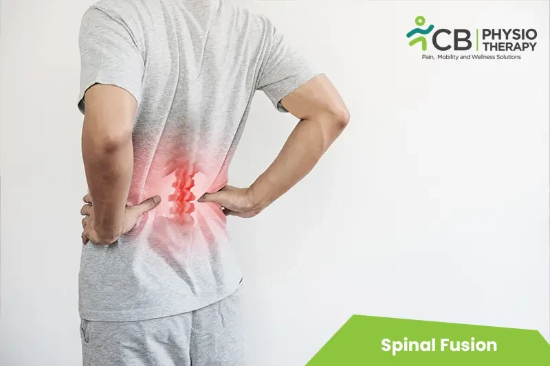
Spinal Fusion Treatment Doctors in Hyderabad

Whether it's a dull ache, a sharp catch, or pain that keeps coming back — we find the root cause of your Spinal Fusion and fix it, at a clinic near you or at home.

Precision Care, Proven Recovery
ACCURATE DIAGNOSIS
ROOT CAUSE ASSESSMENT
COMPREHENSIVE RECOVERY PLAN
MILESTONE TRACKING
GOALS ACHIEVED
STAYING ACTIVE
See how it works ↓
Move pain-free Avoid drugs & surgery

Request received!

We'll call you within 10 minutes to confirm your home visit.We'll call you within 10 minutes to confirm your slot.

Chat with us on WhatsApp

Book in 30 seconds

Visit your nearest CB clinic. Tell us your concern and a preferred time — we'll confirm your slot by call within 10 minutes.

Same-day booking by default. We'll confirm your exact slot by call.
+91
No spam · Your number is safe with us
Prefer to talk now? Call +04045210121
Understanding your spinal fusion

Does this sound like you?

Common triggers include muscle strain, joint stiffness, poor posture and long desk hours. If two or more of these fit, a physiotherapy assessment can identify exactly what's driving it.

  • A dull ache or sharp, burning spinal fusion that keeps returning
  • Discomfort that worsens with movement, lifting or long sitting hours
  • Morning stiffness that eases as you move
  • Pain that limits work, sport or everyday activities
  • Recurring episodes that keep coming back

Most spinal fusion improves without surgery or long-term medication — the key is finding the cause first.

⚠ When to seek urgent medical care

Spinal Fusion rarely signals something serious, but see a doctor immediately if it comes with:

  • Fever, unexplained weight loss, or a history of cancer
  • Numbness or weakness that keeps getting worse
  • Loss of bladder or bowel control
  • Pain after a major fall or accident

Our physios screen for these red flags at every first assessment and refer you to the right doctor if needed.

Assessment-led treatment

How we treat spinal fusion

We start with an AI-based assessment to find the root cause — then follow a clear, four-phase plan.

Phase 1

Ease the pain

Hands-on therapy and advanced modalities to calm pain and inflammation fast.

Phase 2

Restore movement

Mobility work and posture correction so everyday movement feels normal again.

Phase 3

Build strength

Targeted strengthening that protects you — in-clinic or in our physio gym.

Phase 4

Return to activity

Back to work, sport and life — with a home program that prevents relapse.

Your physiotherapist, with Fizo IQ™

Every recovery, planned and proven

One patient record, stage by stage: your physiotherapist pinpoints the diagnosis, finds the root cause and builds your in-clinic plan — while Fizo IQ™, our AI decision-support engine, tracks every milestone until your goals are met and you leave with an exercise plan to stay active.

Physio Expert @CB with Fizo IQ™ — recovery engine
TRACKING
ACCURATE DIAGNOSIS
L4–L5 disc bulge · nerve-root irritation, pinpointed at the first movement assessment
ROOT CAUSE ASSESSMENT
Poor sitting posture, weak core–hip chain · quietly loading that disc
COMPREHENSIVE RECOVERY PLAN
24 clinic sessions · manual therapy + spinal decompression · guided exercise therapy, reviewed weekly
MILESTONE TRACKING
Sitting pain-free ✓ · stairs ✓ · next: the morning 5 km walk
GOALS ACHIEVED
Pain 7 → 3 in six weeks · back to daily life, moving freely
STAYING ACTIVE
A personalized exercise plan, yours to keep · so the pain doesn't come back
Example recovery journey · every plan is built around you
Feeling something?

Symptoms Spinal Fusion addresses

These symptoms respond well to Spinal Fusion — tap one to see how we treat it near you.

Move better

Exercises that help with Spinal Fusion

More exercises →

Make these exercises yours

Get my personalized plan

General guidance only — stop if an exercise increases your pain, and check with your physio before starting a new routine.

Meet your care team

Licensed experts who treat hundreds like you every month

Every CB physio is a qualified BPT/MPT clinician. You don't need to choose one — our lead physiotherapist reviews your case and assigns the specialist best suited to treat it.

Not sure who to see? You don't have to decide. Our lead physiotherapist reviews every case and assigns the physio best suited to it — based on your condition and availability.
Good to know

Spinal Fusion, answered

Spinal fusion is a surgery done to connect two or more vertebrae in the spine, eliminating motion between them. During spinal fusion, a bone or a bone-like material is placed within the space between two spinal vertebrae. Screws, metal plates, and rods may be used to hold the vertebrae together, so they can heal into one solid unit.

Types of Spinal Fusion:

Allograft:

The bone is grafted from any outside source.

Autograft:

The bone is grafted from the patient.
Spinal fusion is used to correct the problems with the small bones in the spine to improve stability, correct a deformity or reduce pain. Other causes may be:


Deformities of the spine.
Herniated disc.
Spinal canal stenosis.
Spinal weakness or instability.
Spondylolisthesis.
Scoliosis.
Fractured vertebra.
Tumour.
Infection.


Pathology:

Spinal fusion is a surgical procedure used to correct problems with the small bones in the spine (vertebrae), and fuse two or more vertebrae so that they heal into one solid bone. Spinal fusion is done to eliminate painful motion or to restore stability to the spine. Spinal fusion eliminates motion between vertebrae and also prevents the stretching of nerves and surrounding ligaments and muscles. Spinal fusion is an option when pain occurs due to movements, such as motion that occurs in a part of the spine that is arthritic or unstable due to injury, the normal aging process, or disease.
X-rays:

X-rays are used to check the bony alignment and bone damage and also show the decreased disk space between the vertebrae.


Computed tomography (CT) scans:

CT scan is used as an adjunctive study to an MRI scan and can also show nerve root compression.


Magnetic resonance imaging (MRI) scans:

MRI scans provide information about the health of the discs as well as the presence of any tumors or compression on the nerves.


Discogram:

Discogram is performed before the spine fusion surgery. It is done by inserting a needle into the disc and injecting dye. If this process causes normal pain, then it is presumed that the specific disc is the anatomic cause of the pain.
Med ica tion: Opioids, nonsteroidal anti-inflammatory drugs, local anesthetics, etc.


Note: Medication should not be taken without the doctor's prescription.
Rest:

Excessive mobility and stress on the tissues should be avoided to enhance healing.


Cryotherapy:

Ice therapy is used to help decrease swelling and pain at the surgical site and is applied for 20 minutes several times a day.


Thermotherapy:

Heat therapy is used to help to improve circulation and relax the muscles.


TENS:

Transcutaneous electrical stimulations may be used to help relieve pain.


Simple range of Motion Exercises:

These exercises help the muscles to maintain the stability of the spine and reduce stress through the surgical site by active stabilization. Range of motion exercises promotes blood circulation, bringing oxygen required for tissue healing and healthy bone growth.


Short walks:

After surgery short walk is recommended for the patients, they should get out of bed and walk the first day after surgery, frequent walking throughout the initial recovery period, and further increase the amount and length of the walks as tolerated.


Stretching Exercises:

Stretching exercises should be done such that the patient should feel the stretch, but not to the point of pain, If pain is felt stretching should be stopped. It promotes flexibility as due to inactivity, range of motion can be lost. Gentle stretching exercises for the core back and abdominals and hip muscles will make all the movements easier. But care should be taken not to do it too aggressively.


Hamstrings and quads Stretching:

Stretching of these muscles should be done slowly with a 30-second hold, 3 times in 2 sets per day. For hamstrings, sit in a chair and straighten one leg in front with toes pointed up and knees straight. Push the belly forward to move into a stretch while keeping the chest high. For the quadriceps, lie on the stomach, and bring the heel toward the buttocks as far as possible.


Nerve stretches:

Nerve stretches should be done in two hours, without holding for long. A nerve stretch is done by lying on the back with legs on the ground, and slowly lifting one leg until a stretch is felt in the back of the thigh. Support the raised leg with hands behind the knee, and pump the ankle while holding the knee still. Or lie on the back, and bend both knees. Slowly straighten one leg and push the heel toward the ceiling until a stretch is felt. Alternate stretching each leg.


Static Stabilization Exercises:

These movements are done without moving the trunk and should be completed by moving arms and legs while avoiding any rocking or arching of the lower trunk.


Pelvic Tilt:

The patient lies on the back of the floor with knees bent and pulling the belly in towards the spine, keeping the lumbar spine stable.


Strengthening Exercises:

More advanced exercises should be added to strengthen the back structures and increase overall fitness. Patients can add more rigor and variety by using an exercise ball or resistance bands.


Mat Exercises:

Abdominal strengthening

The patient lies on the floor with knees bent, curls the trunk by raising the head and one shoulder towards the opposite hip a few inches, then extend the back by alternating the limbs, while on hands and knees, raise one arm and opposite leg, then alternate.


Band Movement:

Strengthen the abdominals and oblique muscles

Resistance bands are commonly used for strength training. Pull with the band, anchored low to the ground, and feet shoulder-width apart, grasp the band and pull from the lower right to the left shoulder.

Exercise Ball Movement:

An exercise ball maximizes the range of motion but staying in control is more important while using an exercise ball. Exercise should be performed until fatigue is evident or control becomes difficult. Each set should last 30 to 60 seconds, 1 set a day is usually recommended.

Ball Marching:

The patient sits on the exercise ball, slowly raising one arm and the opposite leg, reverse.

Another exercise is the patient lies with the stomach on the exercise ball and hands/ arms in front, walk forward on the ball until it rests under the thighs, then raises one leg at a time.

The next option is with the stomach on the exercise ball, and knees on the ground, walk straight out on the hands, but don't let the trunk twist or dip down.


Aerobic Exercises:

Aerobic exercises like stationary biking, stair climbing, elliptical training, or brisk walking for at least 20 minutes helps to increase blood flow and oxygen also burn excess calories, help to maintain weight, and prevent added stress on the back structures and surgical site.

Not all exercises are suitable such as abrupt stops, starts, and changes in direction can put a fusion that is still healing at risk. Jogging or running, sports like basketball or football should be undertaken until a patient has been approved by the surgeon. If there is any shortness of breath, chest pain, or dizziness, then the activity should be immediately stopped.
Ready when you are

Don't let the pain decide your day.

Get assessed today — find the cause, get a plan, and know when you'll feel better.

📞 Call 💬 WhatsApp Book now