
Foot Drop — Symptoms, Causes & Treatment
Also known as Drop Foot. Causes, symptoms and how physiotherapy treats Foot Drop — without surgery or medicines.
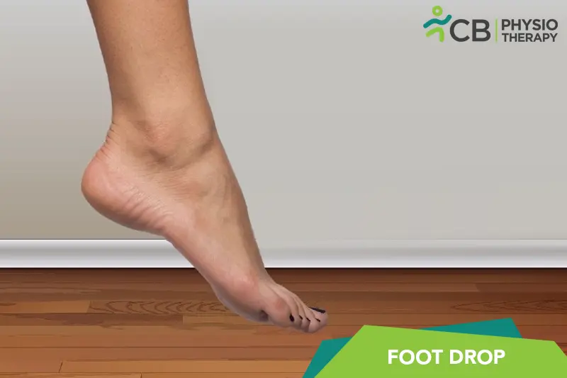
Understanding Foot Drop
Foot Drop (also called Drop Foot) affects people differently, and the right care starts with understanding how it affects you. Its day-to-day impact — on movement, strength, energy and confidence — is where physiotherapy does its work.
Our role in Foot Drop is practical: protect and rebuild movement and daily function with a plan set after a careful assessment, in coordination with your doctor wherever medical management is involved. No two plans look the same, because no two cases behave the same.
Causes & risk factors of Foot Drop
What commonly drives it — your assessment pins down which of these apply to you.
More than one possible driver
Foot Drop can have several contributing factors — a careful history and physical assessment separate them rather than guessing.
Reduced movement and deconditioning
Whatever the trigger, less movement quickly adds weakness and stiffness of its own — usually the most reversible part.
Daily-life load and habits
Work posture, repeated tasks, sleep and stress all shape how a condition behaves day to day.
General health and history
Overall health, previous injuries and existing conditions influence both the picture and the plan — the assessment takes all of it in.
When physiotherapy is the right call
If any of these sound like you, an assessment is the right first step.
- ✓ A new diagnosis of Foot Drop and you want a clear rehabilitation start point
- ✓ Daily tasks, walking or stairs are getting harder than they used to be
- ✓ Strength, stamina or balance has visibly dropped
- ✓ You want a structured clinic + home program with measurable milestones
- ✓ Family members need guidance to support you safely
Your treatment journey
Step by step — from your first assessment to a tracked recovery plan.
A detailed first assessment
History, movement testing and strength checks — a focused session to find what is actually driving your Foot Drop.
A diagnosis you understand
Your physiotherapist explains the findings in plain language — what is affected, why it happened and what that means for you.
Active, tracked treatment
Hands-on therapy, the right modalities and graded exercise — progressed session by session and tracked on Fizo IQ™.
Prevention, built in
The final phase rebuilds strength and habits so the same problem does not come back — with a home plan you keep.
What recovery looks like
- ✓ More independence in daily tasks
- ✓ Better strength, balance and stamina
- ✓ Fewer setbacks, with red flags monitored
- ✓ A program your family understands and can support
- ✓ Progress measured milestone by milestone on Fizo IQ™
When to see a doctor — red flags
How CB Physiotherapy treats Foot Drop
We treat Foot Drop the way we treat everything: assessment first, plan second, treatment third. It sounds obvious, but it is the difference between sessions that fill a slot and sessions that move you towards a goal. Care is delivered by certified physiotherapists at a clinic near you or at home.
Your whole journey is tracked on Fizo IQ™ — the diagnosis, the root cause, the milestones and the goals — so progress is measured, not guessed, and the plan adapts the moment your response calls for it.
Get Foot Drop treatment near you
Certified physiotherapists at a clinic near you or at home — same-day slots, assessment-led care.
Request received!
We'll call you within 10 minutes to confirm your home visit.We'll call you within 10 minutes to confirm your slot.
Chat with us on WhatsAppFoot Drop, answered
1: Nerve Injury: Damage to the peroneal nerve controls the muscles that lift the foot.
2: Muscle Disorders: Conditions such as muscular dystrophy or polio.
3: Brain and Spinal Cord Disorders: Strokes, multiple sclerosis, or cerebral palsy.
4: Herniated Disc: A disc in the spine pressing on nerves.
5: Spinal Stenosis: Narrowing of the spaces within the spine, putting pressure on nerves.
6: Peripheral Neuropathy: Damage to the peripheral nerves, often due to diabetes or alcoholism.
7: Infections: Certain infections affecting the nerves, like Lyme disease or HIV.
8: Tumors or Cysts: Growths that press on nerves.
9: Prolonged Bed Rest or Leg Crossing: Prolonged immobility or pressure on the nerves.
10: Exposure to Toxins: Certain toxins or medications can damage nerves.
11: Trauma: Direct injury to the leg, hip, or spine.
1: Difficulty Lifting the Front Part of the Foot: This is the primary symptom, making it hard to lift the toes and front of the foot off the ground.
2: Dragging the Foot: As a result of the inability to lift the foot, it may drag along the ground when walking.
3: High-Stepping Gait (Steppage Gait): To prevent the foot from dragging, individuals may raise their thigh higher than usual when walking, similar to climbing stairs.
4: Slapping Foot: The foot may slap down onto the floor with each step due to the lack of control.
5: Numbness or Tingling: Some individuals may experience numbness or tingling in the foot or along the shin.
6: Muscle Weakness: There may be noticeable weakness in the muscles of the lower leg and foot.
7: Pain: Pain may be present in the affected area, particularly if there is nerve damage or compression.
8: Difficulty with Balance and Stability: The inability to properly lift and place the foot can lead to problems with balance and an increased risk of falls.
9: Visible Muscle Atrophy: In long-standing cases, there may be a noticeable reduction in muscle mass in the affected leg.
Pathology
The pathology of foot drop involves disruption in the normal function of the nerves and muscles responsible for dorsiflexion, which is the action of lifting the front part of the foot. Overall, the pathology of foot drop involves complex interactions between nerve damage, muscle weakness, and central nervous system dysfunction.
1: Clinical Evaluation:
Medical History: Review of symptoms, medical history, and any recent injuries or illnesses.
2: Physical Examination:
Gait Analysis: Observation of walking patterns to identify high-stepping gait or foot slapping.
Muscle Strength Testing: Assessment of the strength of muscles responsible for dorsiflexion.
Sensation Testing: Checking for numbness or altered sensation in the leg and foot.
Reflex Testing: Evaluating tendon reflexes to assess nerve function.
3: Electromyography (EMG):
Measures electrical activity of muscles to detect abnormalities in muscle function.
Helps identify nerve or muscle disorders affecting the ability to lift the foot.
4: Nerve Conduction Studies (NCS):
Measures the speed and strength of electrical signals traveling through the nerves.
Identifies nerve damage or compression, particularly in the peroneal nerve.
5: Magnetic Resonance Imaging (MRI):
Provides detailed images of soft tissues, nerves, and the spine to detect herniated discs, spinal stenosis, tumors, or other structural abnormalities.
6: Computed Tomography (CT) Scan:
Offers detailed cross-sectional images of bones and soft tissues to identify structural causes of nerve compression.
7: Ultrasound:
Ultrasound helps to visualize nerves and muscles to detect abnormalities or compressions.
8: Blood Tests:
May be used to check for underlying conditions such as diabetes, infections, or autoimmune diseases that can affect nerve function.
9: Lumbar Puncture (Spinal Tap):
In cases of suspected central nervous system disorders, cerebrospinal fluid analysis can help diagnose conditions like multiple sclerosis or infections.
10: Genetic Testing:
If a hereditary neuromuscular disorder is suspected, genetic testing can identify specific mutations associated with conditions like muscular dystrophy.
11: Muscle Biopsy:
Examines muscle tissue for signs of disease or damage.
12: Nerve Biopsy:
Analyses nerve tissue to detect specific patterns of nerve damage.
13: Gait Analysis:
Gait analysis utilizes advanced techniques and equipment to analyze walking patterns in detail.
14: Dynamic Electromyography:
Dynamic electromyography helps assess muscle activity during movement. Each diagnostic technique provides valuable information to help determine the cause of foot drop and guide appropriate treatment strategies.
(Note: Medication should not be taken without the doctor’s prescription.)
Surgery:
Surgeries for foot drop aim to address the underlying cause, improve foot function, and enhance quality of life. Here are some common surgical options:
Tendon Transfer:
Posterior Tibial Tendon Transfer: This surgery involves rerouting the posterior tibial tendon to a position where it can help lift the foot. It’s often used when there is irreversible nerve damage.
Peroneus Longus to Peroneus Brevis Transfer: This procedure transfers the peroneus longus tendon to the peroneus brevis tendon to help lift the foot.
Nerve Decompression:
Nerve Release Surgery: If foot drop is due to nerve compression (e.g., at the knee or lower leg), surgery can release the compressed nerve to restore function.
Ankle Fusion (Arthrodesis):
This procedure fuses the ankle joint in a functional position. It’s usually considered when other treatments have failed, or the condition is severe.
Tendon Lengthening:
If the Achilles tendon is too tight, a procedure to lengthen it can help improve foot function.
Muscle-Tendon Unit (MTU) Shortening:
In some cases, shortening of specific tendons or muscles may be performed to enhance their function and improve foot position.
Heat and Cold Therapy:
Heat therapy also known as thermotherapy and cold therapy also known as cryotherapy helps to manage pain and inflammation.
Functional Electrical Stimulation (FES):
Purpose: To stimulate the peroneal nerve and the muscles responsible for dorsiflexion (lifting the foot).
Application: Electrodes are placed over the nerve or muscle to deliver electrical impulses, helping the foot lift during walking. FES is often used during gait training to improve walking patterns and reduce foot drop.
Transcutaneous Electrical Nerve Stimulation (TENS):
Purpose: Primarily for pain relief.
Application: Electrodes are placed on the skin near the affected area to deliver electrical impulses that interfere with pain signals sent to the brain, helping to alleviate discomfort associated with foot drop.
Neuromuscular Electrical Stimulation (NMES):
Purpose: To strengthen weak muscles and prevent muscle atrophy.
Application: Electrodes are placed on the skin over the affected muscles, and electrical impulses are used to cause muscle contractions. This helps maintain or improve muscle mass and strength.
Interferential Current (IFC) Therapy:
Purpose: Pain relief and reduction of inflammation.
Application: Uses two high-frequency currents that intersect to create a low-frequency current that penetrates deeper into tissues. Electrodes are placed around the affected area to deliver the current.
Iontophoresis:
Purpose: Delivering medication directly to the affected area through the skin.
Application: Uses a mild electrical current to drive anti-inflammatory or pain-relieving medications into the tissue, helping to reduce pain and inflammation.
High-Voltage Pulsed Galvanic Stimulation (HVPGS):
Purpose: To reduce pain and edema, promote wound healing, and stimulate muscle activity.
Application: High-voltage, low-frequency electrical pulses are delivered to the affected area through electrodes.
Russian Stimulation:
Purpose: To enhance muscle strength and re-education.
Application: Uses medium-frequency electrical currents to stimulate muscle contractions. Often used for muscle strengthening and re-training.
Microcurrent Therapy:
Purpose: To promote tissue healing and reduce pain.
Application: Uses very low-intensity electrical currents that mimic the body's natural electrical impulses to stimulate cellular repair and reduce pain.
Strengthening Exercises:
a) Dorsiflexion Exercises: Strengthening the muscles that lift the foot, such as the tibialis anterior.
Ankle Dorsiflexion with Resistance Band: Sitting or lying down, loop a resistance band around the foot and pull the toes toward the body.
b) Plantar Flexion Exercises: Strengthening the calf muscles to provide better support and balance.
c) Heel Raises: Standing and lifting the heels off the ground.
Stretching Exercises:
a) Calf Stretches: To maintain flexibility in the Achilles tendon and prevent tightness.
b) Wall Stretch: Leaning against a wall with one foot forward and one foot back, stretching the back calf.
c) Hamstring Stretches: To ensure the entire lower leg remains flexible.
Balance and Proprioception Training:
Single-Leg Stands: Standing on one leg to improve balance.
Balance Board Exercises: Using a wobble board to challenge stability and proprioception.
Gait Training:
Heel-to-Toe Walking: Practicing walking with a heel-to-toe pattern to improve gait mechanics.
Assisted Walking: Using parallel bars or a therapist’s assistance to practice proper walking techniques.
Treadmill Training: To improve walking endurance and gait pattern, sometimes with body-weight support.
Manual Therapy:
Soft Tissue Mobilization: Massage techniques to reduce muscle tightness and improve circulation.
Joint Mobilization: Techniques to enhance joint flexibility and movement.
Orthotic Devices:
Ankle-Foot Orthoses (AFOs): Braces that support the foot and ankle, holding them in a more functional position and preventing foot drop during walking.
Aquatic Therapy:
Exercising in water to reduce the impact on joints and improve muscle strength.
Functional Training:
Task-Specific Training: Practicing activities of daily living (e.g., stepping over objects, climbing stairs) to improve functional mobility.
Don't let Foot Drop decide your day.
Get assessed today — find the cause, get a plan, and know when you'll feel better.