
Patellar Fracture — Symptoms, Causes & Treatment
Also known as Patellar Fracture. Causes, symptoms and how physiotherapy treats Patellar Fracture — without surgery or medicines.
Understanding Patellar Fracture
Living with Patellar Fracture usually means negotiating with it daily — adjusting how you sit, move, sleep and work around it. It is common; that never makes it something you simply have to live with.
In most cases the problem behaves mechanically: particular movements or positions load the affected structures more than they can currently tolerate. A physiotherapy assessment identifies that mechanism, and treatment targets it directly — most mechanical cases respond well to conservative care, without medicines or surgery.
Causes & risk factors of Patellar Fracture
What commonly drives it — your assessment pins down which of these apply to you.
Sudden spikes in activity
A new sport, a new gym program, a long trek — tissue adapts to gradual change and protests when change is sudden.
Muscle strain and overload
A sudden lift, an awkward movement or simply more load than the tissues were prepared for — the most common trigger.
Posture and long static hours
Desks, driving and screens keep the same structures loaded for hours at a stretch — comfortable at first, aching later.
Joint stiffness and age-related change
Joints that move less start to hurt more; age-related wear is normal, but painful stiffness is not something to simply accept.
Weakness and deconditioning
When supporting muscles lose strength, load shifts to structures that were never meant to carry it.
When physiotherapy is the right call
If any of these sound like you, an assessment is the right first step.
- ✓ Patellar Fracture that keeps returning or hasn't settled within two weeks
- ✓ Pain or stiffness that limits work, sleep or daily activities
- ✓ You want to avoid long-term painkillers — or surgery has been mentioned
- ✓ You've been diagnosed with Patellar Fracture and told to "rest and see"
- ✓ Movement feels restricted, weak or guarded compared to before
Your treatment journey
Step by step — from your first assessment to a tracked recovery plan.
A detailed first assessment
History, movement testing and strength checks — a focused session to find what is actually driving your Patellar Fracture.
A diagnosis you understand
Your physiotherapist explains the findings in plain language — what is affected, why it happened and what that means for you.
Active, tracked treatment
Hands-on therapy, the right modalities and graded exercise — progressed session by session and tracked on Fizo IQ™.
Prevention, built in
The final phase rebuilds strength and habits so the same problem does not come back — with a home plan you keep.
What recovery looks like
- ✓ Care without long-term medicines — at a clinic near you or at home
- ✓ Relief that comes from treating the cause, not masking it
- ✓ A clear diagnosis and a timeline you can plan around
- ✓ Strength and movement rebuilt, not just pain reduced
- ✓ Lower chance of recurrence, with a prevention plan
When to see a doctor — red flags
How CB Physiotherapy treats Patellar Fracture
At CB Physiotherapy, treatment for Patellar Fracture starts with finding its cause: a structured, physiotherapist-led assessment before anything is prescribed. Your plan then combines hands-on therapy, targeted exercise and the right modalities — delivered at a clinic near you or at home, and coordinated with your doctor where medical care is involved.
Every case runs on Fizo IQ™, our recovery engine: diagnosis, root cause, plan, milestones and goals, tracked session by session. You always know where you are in your recovery — and what comes next.
Exercises that help with Patellar Fracture
Make these exercises yours — a CB physio personalizes, doses and tracks your plan week by week.
Get my personalized planGeneral guidance only — stop if an exercise increases your pain, and check with your physio before starting a new routine.
Get Patellar Fracture treatment near you
Certified physiotherapists at a clinic near you or at home — same-day slots, assessment-led care.
Request received!
We'll call you within 10 minutes to confirm your home visit.We'll call you within 10 minutes to confirm your slot.
Chat with us on WhatsAppPatellar Fracture, answered
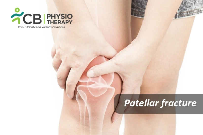
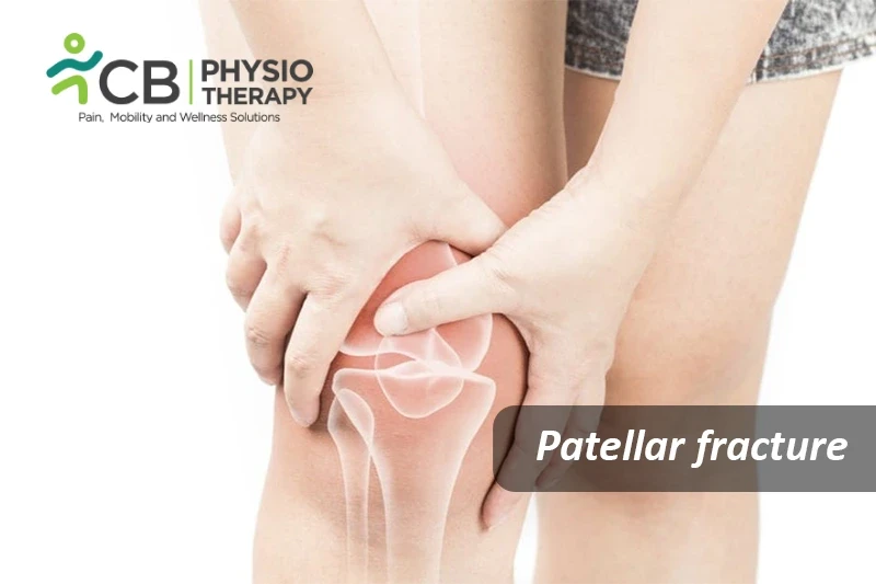
Patella is the small bone that sits at the front of the knee, a crack in this bone is known as a patellar fracture. Patella acts as a shield for the knee joint, covers and protects the knee, and is thus vulnerable to fracture making it difficult or even impossible to straighten the knee or walk.
Types of Patellar fracture
Stable fracture : This type of fracture is a non-displaced fracture. The pieces of bone may remain in contact with each other or be displaced by just a millimeter or two.
Displaced fracture : In a displaced fracture, the broken ends of the bone are separated, normally the smooth joint surface may also be disrupted and thus often requires surgery to put the pieces of bone back together.
Comminuted fracture : In this type of fracture, the bone is displaced into three or more pieces. These fractures may be stable or unstable. In some fractures, the top or the bottom of the patella is broken into several small pieces.
Open fracture: Open fracture involves damage to the soft tissues. Bone fragments come out through the skin and once the skin is broken, there is a higher risk for infection thus requiring immediate treatment to prevent infection.
Closed fracture: Closed fracture involves damage but does not penetrate the skin, there can be severe soft-tissue injury associated with it.
Patellar fractures are most often caused due to:
- Direct fall onto the knee,
- A sharp blow to the knee,
- Head-on vehicle collision if the kneecap is driven into the dashboards,
- Sudden contraction of the quadriceps muscle can pull the patella apart.
The most common symptoms of patellar fracture are pain and swelling, other symptoms include:
- Severe pain in and around the kneecap,
- Swelling and tenderness,
- Bruising,
- Difficulty in extending the leg,
- Difficulty in doing straight-leg-raise,
- Deformed knee,
- Inability to walk.
Pathology:
Patella fractures are caused by trauma, compressive force, or excessive stress to the extensor mechanism, or due to sudden contraction of the quadriceps muscle. Injuries can also be associated with tears of the retinaculum and vastus muscles .
Physical Examination:
Physical examination of the whole limb is done thoroughly. Palpation, flexibility, swelling, range of motion, gait, and overall function and mobility is checked. The knee is also checked for haemarthrosis.
X-rays:
X-rays help to see the images of dense structures, such as bone. X-rays from different angles are done to look for a fracture and the alignment of the bones. X-ray of a patellar fracture shows significant displacement between the broken pieces of bone.
Magnetic resonance imaging (MRI):
Magnetic resonance imaging is done to diagnose the injuries to the tendons and ligaments.
Medication: Non-steroid anti-inflammatory drugs, Opioids, acetaminophen etc.
Note: Medication should not be taken without the doctor's prescription.
Nonsurgical Treatment:
Undisplaced patellar fractures can be treated by wearing a cast or splint to keep the knee straight and prevent motion in the leg. This keeps the broken ends of the bone in the proper position while they heal.
Surgical Treatment:
- In case the pieces of the patellar bone move out of place after injury, then surgery is needed to restore and stabilize the kneecap and allow for the return of function.
- Surgery depends on the type of fracture.
- Open fractures, have an increased risk of infection and thus are scheduled for surgery as soon as possible. During surgery, the cuts from the injury and the surfaces of the bone are cleaned thoroughly and the bone is repaired during the surgery.
- Transverse fracture in which two-part fractures are most often fixed in place using screws, pins, and wires, followed by a tension band. This figure-of-eight band presses the two pieces together.
- This technique is used for fractures that are located near the center of the patella. Another method used to fix the transverse fracture is to secure the bones using small screws and small plates.
- Complete removal of the kneecap is done in case of comminuted fracture.
Cryotherapy is very effective to decrease pain and edema.
Immobilization:
The knee is immobilized in extension either by applying a brace in case of non-surgical treatment or by applying a cast after surgical treatment.
Elevation:
The limb is kept in an extended position and elevation to decrease the swelling.
Ultrasound therapy helps to break the adhesions and decrease pain.
Transcutaneous electrical stimulation (TENS):
Transcutaneous electrical stimulation (TENS) is given to reduce swelling and pain in the knee, also helps to improve muscle recruitment.
Laser therapy (LLLT) accelerates the healing of fractures and enhances callus formation.
Scar massage and mobilization:
Scar massage and mobilization help reduce scar adhesions and thus improve mobility around the scar.
The range is motion exercises are initially done in the inner range these include knee flexion, knee extension, SLR, etc.
Strengthening exercises include isometrics, isotonic exercises for quadriceps, hamstring, adductor, abductor, gluteal and ankle muscles. Resisted exercises with therabands are also given.
Weight Bearing:
Initial weight-bearing exercise is usually limited to gently touching the toe to the floor, with a walker, crutches, or cane. As the injury heals and the muscles strengthen, gradually more weight is put on the leg.
Balance and proprioception exercises:
Balance and proprioception exercises are recommended on a wobble board or a bobath ball.
The patient is made aware that the process of healing depends on age and health, it takes about three to six months to recover from a fractured patella, but in very severe cases it might take longer. The patient is advised to avoid climbing stairs, squatting, kneeling, or other activities that might put a strain on the knee joint.
Don't let Patellar Fracture decide your day.
Get assessed today — find the cause, get a plan, and know when you'll feel better.