Dr. Nayima Tassaduq
Head Of The Clinic · BPT, MPT
See clinics, physiotherapists and prices for your city.
Whether it's a dull ache, a sharp catch, or pain that keeps coming back — we find the root cause of your Posterior Tibial Tendon Dysfunction (pttd) and fix it, at a clinic near you or at home.
Precision Care, Proven RecoveryWe'll call you within 10 minutes to confirm your home visit.We'll call you within 10 minutes to confirm your slot.
Chat with us on WhatsAppCommon triggers include muscle strain, joint stiffness, poor posture and long desk hours. If two or more of these fit, a physiotherapy assessment can identify exactly what's driving it.
Most posterior tibial tendon dysfunction (pttd) improves without surgery or long-term medication — the key is finding the cause first.
Posterior Tibial Tendon Dysfunction (pttd) rarely signals something serious, but see a doctor immediately if it comes with:
Our physios screen for these red flags at every first assessment and refer you to the right doctor if needed.
We start with an AI-based assessment to find the root cause — then follow a clear, four-phase plan.
Hands-on therapy and advanced modalities to calm pain and inflammation fast.
Mobility work and posture correction so everyday movement feels normal again.
Targeted strengthening that protects you — in-clinic or in our physio gym.
Back to work, sport and life — with a home program that prevents relapse.
One patient record, stage by stage: your physiotherapist pinpoints the diagnosis, finds the root cause and builds your in-clinic plan — while Fizo IQ™, our AI decision-support engine, tracks every milestone until your goals are met and you leave with an exercise plan to stay active.
Make these exercises yours — a CB physio personalizes, doses and tracks your plan week by week.
Get my personalized planGeneral guidance only — stop if an exercise increases your pain, and check with your physio before starting a new routine.
Every CB physio is a qualified BPT/MPT clinician. You don't need to choose one — our lead physiotherapist reviews your case and assigns the specialist best suited to treat it.
Head Of The Clinic · BPT, MPT
The same prices you'll see at the clinic — published before you book. Sessions run 30–45 minutes.
Booking a course of sessions? Save 10% when you take a treatment package — and recover with a plan built around your goals.
Ask about packages →Rated 5.0 / 5 across 18 verified patient reviews in Delhi.
“I had a great experience at CB Physiotherapy in Safdarjung, where Dr. Nayima treated my persistent back pain. From the moment I arrived, the clinic had a welcoming atmosphere, and Dr. Nayima’s professionalism stood out. She took the time to thoroughly assess my condition and explained the treatment plan clearly. Over the course of my sessions, I noticed significant improvement in my pain levels and mobility. Dr. Nayima was attentive, skilled, and always ensured I was comfortable during the treatment. I highly recommend CB Physiotherapy for anyone dealing with back pain or other physical issues.”
“Very knowledgeable and gave a great explanation of what was causing me to have such pain in my back. over all treatment was Fantastic.”
“I consulted Dr. Nayima at CB Physiotherapy for spine alignment and fracture pain, and I was quite pleased with the care I received. She developed a tailored treatment plan that included manual therapy and exercises, resulting in great alleviation and improvement. Dr. Nayima possesses expertise, professionalism, and compassion. I strongly suggest her for any spine-related disorders or fractures.”
“I've been going here for just over a month, and dr Nayima has done wonders for my back and neck issue . My only regret is that I did not arrive here sooner. The whole crew is kind and helpful. If you need physical therapy, come here..”
“This place has been fantastic. The staff and therapists are exceptional and extremely informed. I've been to various physical therapy clinics, and this is by far the most useful for my leg and back problems. I would definitely suggest.”
“Recieved treatment from Dr nayima for back and neck problem and i can say that i found relief from pain after couple of sessions. all staff members are very caring.”
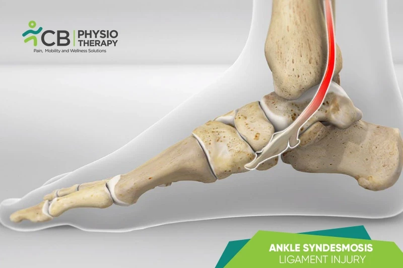
Posterior tibial tendon dysfunction (PTTD) or posterior tibial tendonitis or posterior tibial tendon insufficiency is an issue that causes foot pain. The posterior tibial tendon connects the calf muscle to bones on the inner side of the foot. The main purpose of the tendon is to support the arch on the inside of the foot. It occurs when the posterior tibial tendon swells or gets torn. As a result, the tendon might not provide stability and support for the arch of the foot, resulting in flatfoot. It can be a painful injury that affects the foot and ankle movements, such as walking and running.
There are four stages of posterior tibial tendon dysfunction (PTTD):
Stage I: The tendon is injured but remains intact.
Stage II: The tendon is ruptured or not working properly and the foot is deformed.
Stage III: The foot is significantly deformed including degenerative changes to the cartilage in the back of the foot.
Stage IV: The ankle joint has degenerative changes.
Posterior tibial tendon dysfunction (PTTD) is the most common cause of acquired flatfoot in adults. Causes for PTTD include:
Posterior tibial tendon dysfunction (PTTD) is a painful condition, making certain movements difficult. These movements may include standing, walking, running, or standing on toes, other symptoms include:
Pathology:
PTTD may be due to chronic overpronation or overstretching of the posterior tibialis tendon. Though the posterior tibialis tendon pathology involves the failure of other interosseous ligaments, such as the spring ligament.
Physical examination:
The examiner looks for swelling along the posterior tibial tendon and range of motion in the ankle and foot. Swelling, tenderness, and pain or weakness while moving the foot or ankle are early signs of PTTD. The examiner also looks from behind to look for any changes in its foot structure or shape. The heel may point outward, and the inner arch may rest flat on the ground. The front of the foot may also move away from the body to counterbalance the changes to the heel and inner arch.
Too many toes:
When a normal foot is viewed from behind, only the fifth toe and part or all of the fourth toe are visible on the outside of the foot. But in the case of PTTD, more toes may be visible.
Single-limb heel-rise test:
The patient is asked to stand next to a wall or chair to support the balance. Then asked to raise the unaffected foot off the ground and attempt to lift it onto the toes of the affected foot. With a healthy tendon, an individual should be able to complete eight to 10 heel raises comfortably. In the early stages of PTTD, it may not be possible to complete one single heel raise.
X-rays:
X-rays of the front, back, and sides of the foot provide detailed images of the bones. X-ray helps to rule out arthritis or fallen arches. It also helps to spot joint degeneration in later stages of PTTD.
Magnetic resonance imaging (MRI):
Magnetic resonance imaging (MRI) determines the condition of the tendon and the surrounding muscles.
Computerized tomography scan (CT scan):
A computerized tomography scan (C T scan) creates a 3D image of the soft tissues and bones and provides more detailed images than an x-ray. It also helps to spot arthritis or confirm PTTD.
Ultrasound:
An ultrasound can help to examine the size of the tendon and observe any tendon degeneration or spot fluid in the tissue that surrounds the tendon, which may appear in the early stages of PTTD.
Medication : Non-steroidal anti-inflammatory drugs (NSAIDs) like aspirin, ibuprofen, naproxen, steroid injections, etc.
Note: Medication should not be taken without the doctor's prescription.
PTTD treatment depends on the symptoms. If tendon damage is identified in its earliest stages, then symptoms may be treated by conservative intervention.
Surgery:
Surgery is recommended if the pain does not improve after 6 months of appropriate treatment. The type of surgery depends on where tendonitis is located and the extent of tendon damage. Some of the most commonly done surgeries are:
Rest:
Activities that cause or worsen the pain should be avoided. Low-impact exercises can help to maintain overall health without affecting the tendon. These include bicycling, elliptical training, and swimming.
Cryotherapy or cold therapy can be applied on the most painful area of the posterior tibial tendon for 20 minutes, 3 or 4 times a day to reduce the swelling. It should not be applied directly to the affected part. Place ice over the tendon immediately after completing an exercise, this helps to decrease the swelling around the tendon.
Cast or boot:
A leg cast or walking boot may be used for a few weeks. This allows the tendon to rest and the swelling decreases.
Orthotics:
Orthotics and braces can also be recommended. An orthotic is a shoe insert used for the treatment of a flatfoot. A custom orthotic is required in patients who have moderate to severe changes in the foot. An ankle brace may be used in case of mild to moderate flatfoot. The brace supports the joints of the back of the foot and takes the tension off the tendon. A custom-molded leather brace is recommended for severe flatfoot that is stiff or arthritic. Brace also helps some patients to avoid surgery.
Transcutaneous electrical stimulation (TENS):
Transcutaneous electrical stimulation (TENS) is an electrical modality that can be used to control pain and swelling.
Therapeutic ultrasound can stimulate cell migration, proliferation, and collagen synthesis of tendon cells which can benefit the healing process.
Shockwave therapy is an innovative approach for treating posterior tibialis tendon dysfunction, as proved by various research studies.
Manual therapy uses hands to help improve the ankle moves after posterior tibialis tendon dysfunction (PTTD). After immobilization, the joints of the ankle and toes may be stiff, and thus joint mobilizations may be necessary to improve overall mobility.
Range of motion exercises:
Ankle ROM exercises include pulling the toes and ankle up, pointing the toes and ankle down, moving the foot and ankle inwards, and moving the foot and ankle to the side and away from the body (eversion), these exercises for PTT dysfunction should not hurt.
Stretching exercises:
Calf stretches while standing are a great way to stretch the tendon and the muscles surrounding it. A foam roller can also be used to loosen the calf muscles.
Ankle strengthening exercises add stability to the foot and ankle. This takes the stress and strain from the injured posterior tibialis tendon. Strengthening the ankles with resistance bands is an easy way. Wrap the band around the foot to create resistance while moving the foot. Few exercises that can be done with resistance bands are ankle inversion, ankle eversion, ankle dorsiflexion, and ankle plantarflexion. These exercises should not hurt but should make the ankle and foot feel a little tired. Strengthening the muscles in the hips and knees helps make sure that the foot is in the correct position while moving. Strengthening exercises include squats, lunges, straight leg raises, single-limb heel raises, resistance band exercises, and walking on the toes over a short distance to help prevent injuries.
Balance and proprioception exercises:
Balance and proprioception exercises are a part of a physiotherapy program. Proprioception is the ability to figure out where the body is and how it's moving. Better balance and awareness of the position of the foot and ankle can decrease the stress on the injured tendon. Balance exercises like a single-leg stance progression, standing on a foam pad with one foot and catching a ball, standing on the pad, and squatting down slowly, can be done. Tools like BOSU balls, wobble board, and BAPS board can be used.
Gait training:
Gait training helps to restore normal walking, so gait training may be done during the physiotherapy sessions. The therapist performs specific exercises to help improve the way the patient walks. Assistive devices should also be used so that the progress is proper and safe.
Plyometrics:
Plyometrics are exercises that use the body's ability to jump and land with explosive power. They enable run quickly, change direction, and take the forces on the body while running and jumping. Plyometric exercise can be used as a part of posterior tibial tendon dysfunction rehab. Examples of plyometric exercises are drop jumping, hopping, or jumping in different planes of motion and directions.

The patient is advised not to push through the pain and stop immediately if discomfort is felt. Patients should wear supportive footwear which provides support and is comfortable. Patients should avoid footwear that increases the risk of injury, like high heels, flip-flops, etc.
Get assessed today — find the cause, get a plan, and know when you'll feel better.