Verified
Dr. Sidharth Sharma
Physiotherapist · BPT
See clinics, physiotherapists and prices for your city.
Whether it's a dull ache, a sharp catch, or pain that keeps coming back — we find the root cause of your Temporomandibular Joint (tmj) and fix it, at a clinic near you or at home.
Precision Care, Proven RecoveryWe'll call you within 10 minutes to confirm your home visit.We'll call you within 10 minutes to confirm your slot.
Chat with us on WhatsAppCommon triggers include muscle strain, joint stiffness, poor posture and long desk hours. If two or more of these fit, a physiotherapy assessment can identify exactly what's driving it.
Most temporomandibular joint (tmj) improves without surgery or long-term medication — the key is finding the cause first.
Temporomandibular Joint (tmj) rarely signals something serious, but see a doctor immediately if it comes with:
Our physios screen for these red flags at every first assessment and refer you to the right doctor if needed.
We start with an AI-based assessment to find the root cause — then follow a clear, four-phase plan.
Hands-on therapy and advanced modalities to calm pain and inflammation fast.
Mobility work and posture correction so everyday movement feels normal again.
Targeted strengthening that protects you — in-clinic or in our physio gym.
Back to work, sport and life — with a home program that prevents relapse.
One patient record, stage by stage: your physiotherapist pinpoints the diagnosis, finds the root cause and builds your in-clinic plan — while Fizo IQ™, our AI decision-support engine, tracks every milestone until your goals are met and you leave with an exercise plan to stay active.
Every plan combines hands-on care, advanced modalities and guided exercise — matched to your assessment.
Make these exercises yours — a CB physio personalizes, doses and tracks your plan week by week.
Get my personalized planGeneral guidance only — stop if an exercise increases your pain, and check with your physio before starting a new routine.
Every CB physio is a qualified BPT/MPT clinician. You don't need to choose one — our lead physiotherapist reviews your case and assigns the specialist best suited to treat it.
Verified
Physiotherapist · BPT
Verified
Physiotherapist
Verified
Physiotherapist
Verified
Physiotherapist
Verified
Physiotherapist · BPT
Verified
Physiotherapist
Verified
Physiotherapist · BPT
Rated 5.0 / 5 across 18 verified patient reviews in Faridabad.
“Dr Rahul Kumar is very experienced. I was diagnosed with disc issues and initially I was not even able to move. His treatment helped me recover in a short duration of time.”
“Went for rehab post an elbow dislocation. The place has good facilities and the therapists there are really helpful Dr Rahul Kumar was overseeing me, she was extremely patient and explained things well. The variety of new exercises done helped with my recovery and made the sessions more interesting.”
“Excellent physiotherapist”
“Dr. Sidharth extensive knowledge of physiotherapy was evident. He conducted a thorough assessment of my condition, taking the time to understand my medical history and concerns. His ability to explain the underlying causes of my symptoms in a clear and concise manner helped me grasp the importance of the prescribed treatment plan. he empowered me to actively participate in my own recovery.”
“Much appreciated work in physio”
“Quality treatment!! I will give u 5-stars⭐⭐⭐⭐⭐ for your work...”
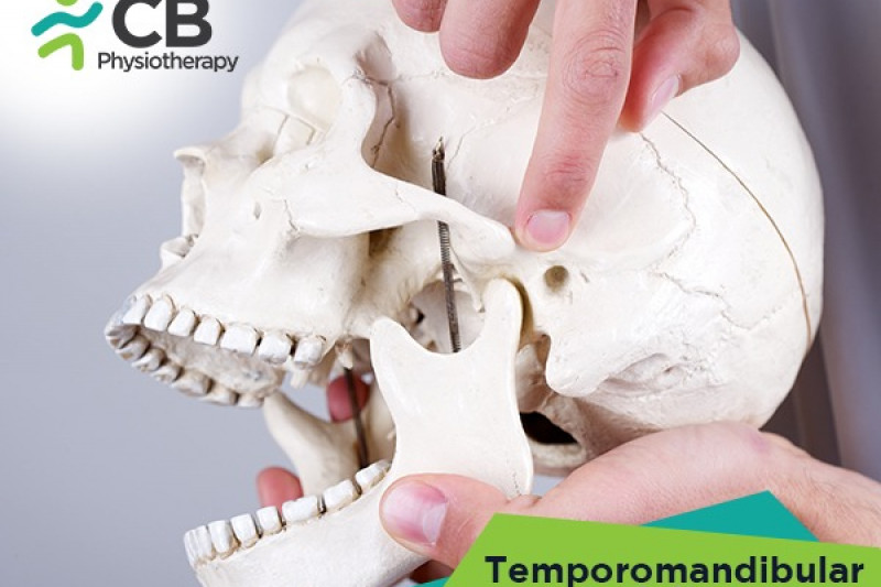
The jaw is also known as TMJ or Temporomandibular joint. These are the most frequently used joints in our body. Without these joints we would be severely hindered, when talking, eating, yawning, kissing, or sucking. It is a synovial joint that connects your jaw to your skull, it is made up of the lower jaw i.e mandible and the socket i.e the temporal bone. There are two temporomandibular joints, one on each side of the jaw. Between the socket and mandible is a disc or menisci.
Sliding, translation or gliding movement occurs in the upper cavity of the Temporomandibular joint, whereas hinge movement or rotation takes place in the lower jaw cavity.
Involvement of this joint results in an inability to open the mouth fully ( approximately 2 inches) with normal side to side gliding and protrusion.
· Pain and tenderness of the masticatory muscles.
· Limited or altered mandibular function (i.e hypermobility, a tendency to protrude the mandible in the initial opening phase).
· Disturbed yawning, eating, biting, shouting, chewing patterns.
· Pain in the temporal area.
· Crepitation or clicking.
· Locking of the jaw.
· Muscle spasm.
· Old injures or trauma.
· Postural dysfunction with a forward head posture.
· Dental problems, malocclusion, decreased vertical dimension of the bite.
· Hindered joint mechanics, dislocation of the condylar head, subluxation of the menisci (disc), or inflammation, asymmetric joint forces, immobilization after jaw fracture or any surgery, for years with eventual adaptation failure, maybe the reason for the present complaint of TMD.
· Mouth breathers in which the jaw position and the posture is affected indirectly.
· Inflammation or muscle spasm in fascial muscles, emotional tension, postural stress syndrome, faulty joint mechanics.
· Excessive stresses such a biting or chewing on large pieces of hard food may also traumatize the joints.
· Osteoarthritis , skeletal limitations and reduced vertical dimensions.
The effective management of Temporomandibular disorders requires first of all a diagnosis based on a complete history and thorough physical examination.
The following tests can aid in determining the intracapsular pathological conditions:
· Dynamic loading of one joint:
Ask the patient to forcefully bite the cotton roll or a tongue blade on one side. This procedure loads the contralateral temporomandibular joint and thus elicits pain.
· Posterior compression or loading of both the Temporomandibular joints :
Hold the mandible with both the hands placing the thumb on the distal molars with the fingers beneath the mandible. The mandible is then tipped down and back to compress the joints. The mandible will also be moved forward or backward to localize tenderness in the posterior or inferior parts of the joint.
· Unloading (distraction) or caudal traction:
Using the same hand positioning as mentioned above, the distraction of both the joints is performed at the same time or caudal traction of each joint is performed. The incisors are not aligned, with the mandible being deviated to one side. To check the relation of this deformity to the patient's symptoms, the therapist passively attempts to correct the deformity. If the deviation is a protective deformity, then the test will increase the pain.
· Plain Film Radiography
On the anteroposterior view, the examiner should look for condylar shape and normal contours. On the lateral view, the examiner should look for the condylar shape and contours, the position of condylar heads in the opened and closed positions, amount of the condylar movement (closed versus open) and the relation of temporomandibular joint to other bony structures of the skull and cervical spine.
· Magnetic Resonance Imaging
This technique is used to differentiate the soft tissue of the joint, the disc from the structures.
· Roentgenography and electromyography ,
Electromyography can reveal how a muscle acts at any point during mandibular movements and procedures. Some researchers believe that electromyography is more reliable diagnostic tool than roentgenography for temporomandibular joints.

The management depends on the cause of the syndrome. In cases in which joint dysfunction, muscle imbalance, or posture are the sources of the problem, intervention with therapeutic exercise can directly address the problems but in other cases, we might need dental or psychological support.
A complete evaluation is necessary before initiating any treatment.
Medication:
Anti – Inflammatory drug, Analgesics to relieve pain, Muscle relaxants, Sedatives
NOTE : Medication not to be taken without the prescription of the doctor.
Cryotherapy on the area of pain for 15 – 20 minutes to reduce inflammation
Thermotherapy for 15 – 20 minutes to increase the blood circulation and to relax the involved muscles
TENS Therapy are electrical stimulations using electric current at different frequencies for the management of pain by sending signals to the brain and offering short term relief.
Ultrasound circulation therapy provides deep heat into the muscles and joints hence increasing the, causing relaxation in the muscles hence reducing pain.
Soft tissue techniques :
· Friction massage to the capsule of the joint.
· Kneading or stroking techniques interiorly to inhibit pain (to the insertion of the temporalis and medial and lateral pterygoid musculature).
· Deep pressure massage.
· Connective tissue massage.
· Muscle energy or post isometric relation techniques.
· Stretching techniques.
For Facial muscles, Tongue proprioception and Tongue control:
· Place the tip of the tongue on the hard palate behind infront teeth and draw circles or letters on the palate.
· Place the tip of the tongue on the hard palate and blow air out to vibrate the tongue making rrrr sound.
· Fill the cheeks with air, mouth closed, then let the air out in a puff.
· Make a clicking sound with the tongue on the roof of the mouth. When doing so, the jaw drops open quickly and returns with the teeth slightly apart usually, the tongue rests on the hard palate behind the front teeth. This is the resting position of the jaw and is also the first step in teaching relaxation exercises.
Jaw muscles
· Starting with the resting position. The lips are closed, the teeth slightly apart, usually the tongue rests on the hard palate behind the front teeth. The patient should breathe in and out slowly through the nose, using diaphragmatic breathing.
· Opening and closing the jaw through the first half of the ROM. With the tongue on the roof of the mouth, the patient opens the mouth, trying to keep the chin in the midline (in front of the mirror). The patient is also taught to lightly palpate the lateral pole of each condyle of the mandible bilaterally and to attempt to maintain symmetry between movement of the two sides when opening and closing the mouth.
· Gold fish exercises half mouth:
Keep to the tongue on the hard palate of the mouth. Place one finger on each joint. Place the other index finger on your chin and then let the mandible (lower jaw) partially drop down and back with help of the index finger.
· Gold fish exercises full mouth:
Keep to the tongue on the hard palate of the mouth. Place one finger on each joint.
let the mandible ( lower jaw) fully drop down and back to bring the chin to the throat.
· Chin tucks
Make a double chin by bringing your chin straight back, keep the head straight up without any bending forward or behind.
Tongue blades can be used for home treatment both as a static stretch and for a briefer period soft active and passive stretch. However, tongue blades must be used with great caution since the too vigorous application may cause injury. Even a tapered cork, which is approx. 15 mm at its narrow end and 30 mm at its widest end, is gradually put into the patient's mouth( small end first) until the jaw separates’. Once the jaw begins to relax, the cork can be placed farther into the mouth, thus opening the jaw wider and wider. This technique can be used for 30 seconds every 2 hours. Yawning is also recommended as a home program.
Most important step is to educate the patient regarding the functioning of the joints, the reason for their symptoms and the means of removing those symptoms. Patient should be advised about the wide opening, excessive mouth yawning, biting off large mouthfuls of hard food instead eat soft food and cut the food into small pieces, habitual protrusion, diurnal clenching should be explained. The methods to achieve muscle relaxation and abolish well-established patterns of inappropriate muscular activity and methods to acquire new ones are important means of eliminating symptoms
Get assessed today — find the cause, get a plan, and know when you'll feel better.