Verified
Dr. Noureen Begum
Physiotherapist · BPT
See clinics, physiotherapists and prices for your city.
Whether it's a dull ache, a sharp catch, or pain that keeps coming back — we find the root cause of your Ankle Syndesmosis Ligament Injury and fix it, at a clinic near you or at home.
Precision Care, Proven RecoveryWe'll call you within 10 minutes to confirm your home visit.We'll call you within 10 minutes to confirm your slot.
Chat with us on WhatsAppCommon triggers include muscle strain, joint stiffness, poor posture and long desk hours. If two or more of these fit, a physiotherapy assessment can identify exactly what's driving it.
Most ankle syndesmosis ligament injury improves without surgery or long-term medication — the key is finding the cause first.
Ankle Syndesmosis Ligament Injury rarely signals something serious, but see a doctor immediately if it comes with:
Our physios screen for these red flags at every first assessment and refer you to the right doctor if needed.
We start with an AI-based assessment to find the root cause — then follow a clear, four-phase plan.
Hands-on therapy and advanced modalities to calm pain and inflammation fast.
Mobility work and posture correction so everyday movement feels normal again.
Targeted strengthening that protects you — in-clinic or in our physio gym.
Back to work, sport and life — with a home program that prevents relapse.
One patient record, stage by stage: your physiotherapist pinpoints the diagnosis, finds the root cause and builds your in-clinic plan — while Fizo IQ™, our AI decision-support engine, tracks every milestone until your goals are met and you leave with an exercise plan to stay active.
These symptoms respond well to Ankle Syndesmosis Ligament Injury — tap one to see how we treat it near you.
Make these exercises yours — a CB physio personalizes, doses and tracks your plan week by week.
Get my personalized planGeneral guidance only — stop if an exercise increases your pain, and check with your physio before starting a new routine.
Every CB physio is a qualified BPT/MPT clinician. You don't need to choose one — our lead physiotherapist reviews your case and assigns the specialist best suited to treat it.
Verified
Physiotherapist · BPT
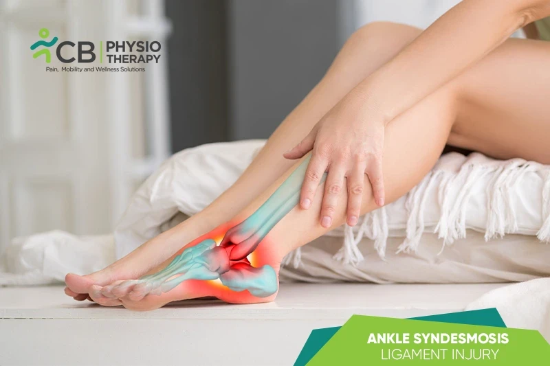
Ankle syndesmosis ligament injury or high ankle sprain is also known as tibiofibular syndesmosis, it is the injury of a ligament that holds the fibrous joint together. It's located near the ankle joint, between the tibia (shinbone), and the distal fibula (outside leg bone). The ligament acts as a shock absorber, providing stability and support for the ankle, and also aligns the tibia and fibula and preventing them from spreading too far apart.
It is made of several ligaments. The primary ones are:
Ankle syndesmosis ligament injury can occur due to many reasons, few causes of the injury can be:
Generally, ankle syndesmosis ligament injuries don't swell or bruise as much as other ankle sprains. Symptoms can vary depending on the severity of the injury:
Pathology:
Ankle syndesmosis ligament injury occurs due to maximal tension when the ankle is fully dorsiflexed or fully plantarflexed or external rotation of the foot on the leg causes the talus to press against the lateral malleolus. Excessive force at the syndesmosis can sprain or rupture the anterior and posterior tibiofibular ligaments, tear the deltoid ligament, force the talus to push the fibula laterally, and damage the lateral ankle ligaments, and also disrupt the ankle mortise and fibular stability.
Physical examination:
Physical examination may be painful, or uncomfortable. The examiner squeezes and manipulates the leg and foot to see how well he/she can be flexed, rotated, and whether they can bear weight.
Squeeze test:
The examiner tries to separate the tibia and fibula and identifies a fibular fracture or syndesmosis sprain. This is performed by squeezing the tibia and fibula together above the injury. Pain is reproduced along the fibular shaft, if it's a fibular fracture and the distal tibiofibular joint for syndesmosis sprain.
X-ray:
X-ray is required to determine if there are one or more broken bones.
Magnetic resonance imaging (MRI):
Magnetic resonance imaging (MRI) can help to detect tears and injuries to ligaments and tendons.
CT scan:
CT scans take X-rays from different angles and combine them to make 3-D images. CT scans are done to reveal more detailed images of the bones of the joint.
Ultrasound:
Ultrasound uses sound waves to produce images that may help the doctor to check the condition of a ligament or tendon when the foot is in different positions.
Medication: Non-steroidal anti-inflammatory drugs, Analgesics, etc.
Note: Medication should not be taken without the doctor's prescription.
Surgery:
Surgery is recommended in higher grade injuries if instability and widening between the two shin bones are significant. Pins and screws may be placed in place to allow the ligament to have a better healing capacity.
Rest:
Rest is recommended to limit any movement that causes pain. This may include non-weight bearing in a brace with crutches.
Cryotherapy or ice therapy is a simple and effective modality used to reduce pain and swelling. Ice is applied for 20-30 minutes.
Compression:
Compression bandaging is recommended to support the injured soft tissue and reduce excessive inflammation.
Elevation:
The affected leg is kept in elevation above the heart to assist gravity to reduce excessive swelling around the ankle.
Ankle support:
An ankle brace can be used to protect the damaged ligament, it provides protection and stability.
Kinesio taping helps to provide mechanical support for an unstable ankle joint.
Transcutaneous Electrical Stimulation:
Transcutaneous Electrical Stimulation is found to be effective to decrease swelling, spasms, and pain.
Therapeutic Ultrasound:
Therapeutic ultrasound decreases pain, and inflammation improves function, and helps to enhance the healing process.
Manual therapy includes soft tissue massage, lymphatic drainage, talocrural distraction in the neutral position, and anterior to posterior talocrural glides. Manual therapy helps in pain reduction, reduces stiffness, and increases functionality.
Range of Motion exercises:
Range of motion exercises are done to increase joint movement and functionality. Simple range of motion exercises can be done to improve joint movement. Passive range of motion exercises for dorsiflexion can be performed in both weight-bearing and non-weight-bearing positions.
Muscle Strengthening exercises:
Calf, ankle, and foot muscles require strengthening after a high ankle sprain. Strengthening exercises provide normal dynamic ankle control and function. Strength and power should be gradually progressed from non-weight bearing to partial and then full weight-bearing and resistance-loaded exercises.
Stretching exercises:
Stretching exercises like standing stretch, seated dorsiflexion, stretch with theraband, double heel raise progressing to single heel raise, and dorsiflexion stretching on a step stool can be used to increase the flexibility of the muscles.
Proprioception and Agility exercises:
Balance and proprioception are adversely affected by a high ankle sprain. To prevent the re-injury, the patient should return to sport, the physiotherapist guides the patient through the exercises to improve the performance. Depending on the type of sport or lifestyle, speed, agility, proprioception, and power program are customized to prepare the patient for light sport-specific training. Ankle proprioception, postural reflexes, and balance reeducation are also done.
Plyometric Training;
Exercise training is progressed to jogging, cycling, agility, jumping, and sport-specific drills involving high-intensity exercises.

The patient is advised to practice foot and ankle functions with a brace during high-intensity exercises for protection and support.
Get assessed today — find the cause, get a plan, and know when you'll feel better.