Open today · 8 AM–8 PM · Same-day slots
CB Physiotherapy
Same-day appointments

Ankle Syndesmosis Ligament Injury Treatment Doctors in Noida

Whether it's a dull ache, a sharp catch, or pain that keeps coming back — we find the root cause of your Ankle Syndesmosis Ligament Injury and fix it, at a clinic near you or at home.

Precision Care, Proven Recovery
ACCURATE DIAGNOSIS
ROOT CAUSE ASSESSMENT
COMPREHENSIVE RECOVERY PLAN
MILESTONE TRACKING
GOALS ACHIEVED
STAYING ACTIVE
See how it works ↓
Move pain-free Avoid drugs & surgery
5.0 / 5 · 31 Google reviews Verified on Google

Request received!

We'll call you within 10 minutes to confirm your home visit.We'll call you within 10 minutes to confirm your slot.

Chat with us on WhatsApp

Book in 30 seconds

Visit your nearest CB clinic. Tell us your concern and a preferred time — we'll confirm your slot by call within 10 minutes.

Same-day booking by default. We'll confirm your exact slot by call.
+91
No spam · Your number is safe with us
Prefer to talk now? Call +09821966696
Understanding your ankle syndesmosis ligament injury

Does this sound like you?

Common triggers include muscle strain, joint stiffness, poor posture and long desk hours. If two or more of these fit, a physiotherapy assessment can identify exactly what's driving it.

  • A dull ache or sharp, burning ankle syndesmosis ligament injury that keeps returning
  • Discomfort that worsens with movement, lifting or long sitting hours
  • Morning stiffness that eases as you move
  • Pain that limits work, sport or everyday activities
  • Recurring episodes that keep coming back

Most ankle syndesmosis ligament injury improves without surgery or long-term medication — the key is finding the cause first.

⚠ When to seek urgent medical care

Ankle Syndesmosis Ligament Injury rarely signals something serious, but see a doctor immediately if it comes with:

  • Fever, unexplained weight loss, or a history of cancer
  • Numbness or weakness that keeps getting worse
  • Loss of bladder or bowel control
  • Pain after a major fall or accident

Our physios screen for these red flags at every first assessment and refer you to the right doctor if needed.

Assessment-led treatment

How we treat ankle syndesmosis ligament injury

We start with an AI-based assessment to find the root cause — then follow a clear, four-phase plan.

Phase 1

Ease the pain

Hands-on therapy and advanced modalities to calm pain and inflammation fast.

Phase 2

Restore movement

Mobility work and posture correction so everyday movement feels normal again.

Phase 3

Build strength

Targeted strengthening that protects you — in-clinic or in our physio gym.

Phase 4

Return to activity

Back to work, sport and life — with a home program that prevents relapse.

Your physiotherapist, with Fizo IQ™

Every recovery, planned and proven

One patient record, stage by stage: your physiotherapist pinpoints the diagnosis, finds the root cause and builds your in-clinic plan — while Fizo IQ™, our AI decision-support engine, tracks every milestone until your goals are met and you leave with an exercise plan to stay active.

Physio Expert @CB with Fizo IQ™ — recovery engine
TRACKING
ACCURATE DIAGNOSIS
L4–L5 disc bulge · nerve-root irritation, pinpointed at the first movement assessment
ROOT CAUSE ASSESSMENT
Poor sitting posture, weak core–hip chain · quietly loading that disc
COMPREHENSIVE RECOVERY PLAN
24 clinic sessions · manual therapy + spinal decompression · guided exercise therapy, reviewed weekly
MILESTONE TRACKING
Sitting pain-free ✓ · stairs ✓ · next: the morning 5 km walk
GOALS ACHIEVED
Pain 7 → 3 in six weeks · back to daily life, moving freely
STAYING ACTIVE
A personalized exercise plan, yours to keep · so the pain doesn't come back
Example recovery journey · every plan is built around you
Feeling something?

Symptoms Ankle Syndesmosis Ligament Injury addresses

These symptoms respond well to Ankle Syndesmosis Ligament Injury — tap one to see how we treat it near you.

Move better

Exercises that help with Ankle Syndesmosis Ligament Injury

More exercises →

Make these exercises yours

Get my personalized plan

General guidance only — stop if an exercise increases your pain, and check with your physio before starting a new routine.

Meet your care team

Licensed experts who treat hundreds like you every month

Every CB physio is a qualified BPT/MPT clinician. You don't need to choose one — our lead physiotherapist reviews your case and assigns the specialist best suited to treat it.

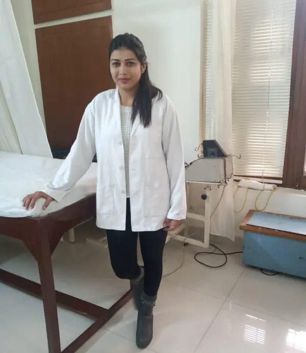
Dr. Vidhi, physiotherapist in Noida Extention Verified

Dr. Vidhi

Physiotherapist · BPT

5.0 (9)
Noida Extention
5 yrs experience
Ortho Spine Sports Physiotherapy Center
Dr. Deepika, physiotherapist in Greater Noida Verified

Dr. Deepika

Physiotherapist

5.0 (1)
Greater Noida
4 yrs experience
Ortho Spine Sports Physiotherapy Center
Not sure who to see? You don't have to decide. Our lead physiotherapist reviews every case and assigns the physio best suited to it — based on your condition and availability.
Patient stories

Ankle Syndesmosis Ligament Injury recoveries, verified on Google

Rated 5.0 / 5 across 31 verified patient reviews in Noida.

Verified

“Dr kasim listened well to my situation, was very quick to diagnose me and gave very clear guidelines on how I should manage my condition”

AB
Aahil Verified patient · Treated at CB · 3 weeks ago
Verified

“I recently had the opportunity to experience physiotherapy at home, and I must say it was an outstanding and convenient experience. One of the biggest advantages of receiving physiotherapy at home was the convenience it offered. Instead of traveling to a clinic or hospital, the physiotherapists came to my doorstep, saving me time and energy. This personalized approach allowed me to receive treatment in the comfort of my own home, which greatly contributed to my overall well-being and recovery. Thanks Dr. kasim for the services.”

RG
Rakhi Gupta Verified patient · Treated at CB · 3 months ago
Verified

“I wholeheartedly recommend Dr. Kasim to anyone seeking treatment for a shoulder issue. His outstanding care, expertise, and compassionate approach make him an exceptional physiotherapist. Under his guidance, I experienced remarkable progress and successful recovery. Thank you, Dr. Kasim, for your exceptional care and dedication to helping your patients regain their shoulder health.”

AH
Anwer Hussain Verified patient · Treated at CB · 3 months ago
Verified

“Dr. Vidhi's approach is highly professional and patient-centered. She took the time to thoroughly assess my condition, asking detailed questions and actively listening to my concerns. She ensured that I fully understood my diagnosis, the treatment plan, and the expected outcomes. . Her dedication to providing evidence-based care was evident. thanks for treated me so well..god bless.”

RK
Rakulpreet Kaur Verified patient · Treated at CB · 3 months ago
Verified

“Thank you sir for providing home physiotherapy treatment. its wonderful to have session with you. 🙏🙏”

SL
Sham Lal Verified patient · Treated at CB · 3 months ago
Verified

“Hats off to you, ma'am. I am very satisfied with the treatment I had for body posture. Every session was great; it made me feel comfortable.👍👍”

AV
Anjali Verma Verified patient · Treated at CB · 3 months ago
Good to know

Ankle Syndesmosis Ligament Injury, answered

Ankle syndesmosis ligament injury or high ankle sprain is also known as tibiofibular syndesmosis, it is the injury of a ligament that holds the fibrous joint together. It's located near the ankle joint, between the tibia (shinbone), and the distal fibula (outside leg bone). The ligament acts as a shock absorber, providing stability and support for the ankle, and also aligns the tibia and fibula and preventing them from spreading too far apart.

It is made of several ligaments. The primary ones are:


  • Anterior inferior tibiofibular ligament
  • Posterior inferior tibiofibular ligament
  • Interosseous ligament
  • Transverse tibiofibular ligament.

Ankle syndesmosis ligament injury can occur due to many reasons, few causes of the injury can be:


  • Tripping over something,
  • Leg rotated internally.
  • External rotation of the talus (bone),
  • Sudden twisting motion,
  • High energy blow to the firmly planted foot,
  • Sports like football, rugby, downhill skiing, hockey,
  • Planting the foot in place while the ankle is forced to rotate outwardly,
  • Blow to the outer side of the ankle.

Generally, ankle syndesmosis ligament injuries don't swell or bruise as much as other ankle sprains. Symptoms can vary depending on the severity of the injury:


  • Pain above the ankle,
  • Pain radiating up to the leg,
  • Tenderness to touch,
  • Pain aggravated while walking,
  • Pain when the foot is flexed or rotated,
  • Inability to raise the calf,
  • Inability to bear full weight.
  • Often accompanied by injuries to bones, ligaments, or tendons.

Pathology:

Ankle syndesmosis ligament injury occurs due to maximal tension when the ankle is fully dorsiflexed or fully plantarflexed or external rotation of the foot on the leg causes the talus to press against the lateral malleolus. Excessive force at the syndesmosis can sprain or rupture the anterior and posterior tibiofibular ligaments, tear the deltoid ligament, force the talus to push the fibula laterally, and damage the lateral ankle ligaments, and also disrupt the ankle mortise and fibular stability.

Physical examination:

Physical examination may be painful, or uncomfortable. The examiner squeezes and manipulates the leg and foot to see how well he/she can be flexed, rotated, and whether they can bear weight.

Squeeze test:

The examiner tries to separate the tibia and fibula and identifies a fibular fracture or syndesmosis sprain. This is performed by squeezing the tibia and fibula together above the injury. Pain is reproduced along the fibular shaft, if it's a fibular fracture and the distal tibiofibular joint for syndesmosis sprain.

X-ray:

X-ray is required to determine if there are one or more broken bones.

Magnetic resonance imaging (MRI):

Magnetic resonance imaging (MRI) can help to detect tears and injuries to ligaments and tendons.

CT scan:

CT scans take X-rays from different angles and combine them to make 3-D images. CT scans are done to reveal more detailed images of the bones of the joint.

Ultrasound:

Ultrasound uses sound waves to produce images that may help the doctor to check the condition of a ligament or tendon when the foot is in different positions.

Medication: Non-steroidal anti-inflammatory drugs, Analgesics, etc.

Note: Medication should not be taken without the doctor's prescription.

Surgery:

Surgery is recommended in higher grade injuries if instability and widening between the two shin bones are significant. Pins and screws may be placed in place to allow the ligament to have a better healing capacity.

Rest:

Rest is recommended to limit any movement that causes pain. This may include non-weight bearing in a brace with crutches.

Cryotherapy:

Cryotherapy or ice therapy is a simple and effective modality used to reduce pain and swelling. Ice is applied for 20-30 minutes.

Compression:

Compression bandaging is recommended to support the injured soft tissue and reduce excessive inflammation.

Elevation:

The affected leg is kept in elevation above the heart to assist gravity to reduce excessive swelling around the ankle.

Ankle support:

An ankle brace can be used to protect the damaged ligament, it provides protection and stability.

Kinesio Taping:

Kinesio taping helps to provide mechanical support for an unstable ankle joint.

Transcutaneous Electrical Stimulation:

Transcutaneous Electrical Stimulation is found to be effective to decrease swelling, spasms, and pain.

Therapeutic Ultrasound:

Therapeutic ultrasound decreases pain, and inflammation improves function, and helps to enhance the healing process.

Manual Therapy:

Manual therapy includes soft tissue massage, lymphatic drainage, talocrural distraction in the neutral position, and anterior to posterior talocrural glides. Manual therapy helps in pain reduction, reduces stiffness, and increases functionality.

Range of Motion exercises:

Range of motion exercises are done to increase joint movement and functionality. Simple range of motion exercises can be done to improve joint movement. Passive range of motion exercises for dorsiflexion can be performed in both weight-bearing and non-weight-bearing positions.

Muscle Strengthening exercises:

Calf, ankle, and foot muscles require strengthening after a high ankle sprain. Strengthening exercises provide normal dynamic ankle control and function. Strength and power should be gradually progressed from non-weight bearing to partial and then full weight-bearing and resistance-loaded exercises.

Stretching exercises:

Stretching exercises like standing stretch, seated dorsiflexion, stretch with theraband, double heel raise progressing to single heel raise, and dorsiflexion stretching on a step stool can be used to increase the flexibility of the muscles.

Proprioception and Agility exercises:

Balance and proprioception are adversely affected by a high ankle sprain. To prevent the re-injury, the patient should return to sport, the physiotherapist guides the patient through the exercises to improve the performance. Depending on the type of sport or lifestyle, speed, agility, proprioception, and power program are customized to prepare the patient for light sport-specific training. Ankle proprioception, postural reflexes, and balance reeducation are also done.

Plyometric Training;

Exercise training is progressed to jogging, cycling, agility, jumping, and sport-specific drills involving high-intensity exercises.

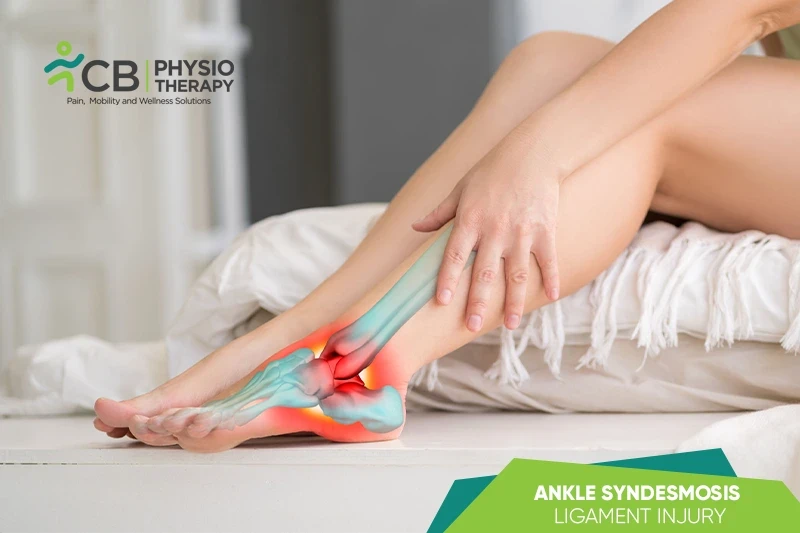
The patient is advised to practice foot and ankle functions with a brace during high-intensity exercises for protection and support.

Ready when you are

Don't let the pain decide your day.

Get assessed today — find the cause, get a plan, and know when you'll feel better.

📞 Call 💬 WhatsApp Book now