
Osgood-schlatter Disease — Symptoms, Causes & Treatment
Also known as Osteochondrosis. Causes, symptoms and how physiotherapy treats Osgood-schlatter Disease — without surgery or medicines.
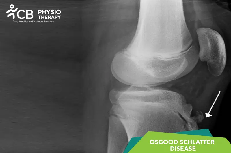
Understanding Osgood-schlatter Disease
Osgood-schlatter Disease (also called Osteochondrosis) affects people differently, and the right care starts with understanding how it affects you. Its day-to-day impact — on movement, strength, energy and confidence — is where physiotherapy does its work.
Our role in Osgood-schlatter Disease is practical: protect and rebuild movement and daily function with a plan set after a careful assessment, in coordination with your doctor wherever medical management is involved. No two plans look the same, because no two cases behave the same.
Causes & risk factors of Osgood-schlatter Disease
What commonly drives it — your assessment pins down which of these apply to you.
More than one possible driver
Osgood-schlatter Disease can have several contributing factors — a careful history and physical assessment separate them rather than guessing.
Reduced movement and deconditioning
Whatever the trigger, less movement quickly adds weakness and stiffness of its own — usually the most reversible part.
Daily-life load and habits
Work posture, repeated tasks, sleep and stress all shape how a condition behaves day to day.
General health and history
Overall health, previous injuries and existing conditions influence both the picture and the plan — the assessment takes all of it in.
When physiotherapy is the right call
If any of these sound like you, an assessment is the right first step.
- ✓ A new diagnosis of Osgood-schlatter Disease and you want a clear rehabilitation start point
- ✓ Daily tasks, walking or stairs are getting harder than they used to be
- ✓ Strength, stamina or balance has visibly dropped
- ✓ You want a structured clinic + home program with measurable milestones
- ✓ Family members need guidance to support you safely
Your treatment journey
Step by step — from your first assessment to a tracked recovery plan.
A detailed first assessment
History, movement testing and strength checks — a focused session to find what is actually driving your Osgood-schlatter Disease.
A diagnosis you understand
Your physiotherapist explains the findings in plain language — what is affected, why it happened and what that means for you.
Active, tracked treatment
Hands-on therapy, the right modalities and graded exercise — progressed session by session and tracked on Fizo IQ™.
Prevention, built in
The final phase rebuilds strength and habits so the same problem does not come back — with a home plan you keep.
What recovery looks like
- ✓ More independence in daily tasks
- ✓ Better strength, balance and stamina
- ✓ Fewer setbacks, with red flags monitored
- ✓ A program your family understands and can support
- ✓ Progress measured milestone by milestone on Fizo IQ™
When to see a doctor — red flags
How CB Physiotherapy treats Osgood-schlatter Disease
At CB Physiotherapy, treatment for Osgood-schlatter Disease starts with finding its cause: a structured, physiotherapist-led assessment before anything is prescribed. Your plan then combines hands-on therapy, targeted exercise and the right modalities — delivered at a clinic near you or at home, and coordinated with your doctor where medical care is involved.
Every case runs on Fizo IQ™, our recovery engine: diagnosis, root cause, plan, milestones and goals, tracked session by session. You always know where you are in your recovery — and what comes next.
Get Osgood-schlatter Disease treatment near you
Certified physiotherapists at a clinic near you or at home — same-day slots, assessment-led care.
Request received!
We'll call you within 10 minutes to confirm your home visit.We'll call you within 10 minutes to confirm your slot.
Chat with us on WhatsAppOsgood-schlatter Disease, answered
1: Rapid Growth Spurts: During adolescence, bones, muscles, and tendons grow at different rates. This mismatch can increase tension at the site where the patellar tendon attaches to the tibia, leading to inflammation and pain.
2: Repetitive Stress from Sports: Activities involving running, jumping, and sudden changes in direction (common in sports like basketball, soccer, gymnastics, and track) put repeated stress on the patellar tendon. This repetitive pulling can cause the tendon to pull away slightly from the tibia, leading to inflammation and swelling.
3: Overuse: Continuous activity without adequate rest can exacerbate the stress on the growth plate at the tibial tuberosity, increasing the risk of developing Osgood-Schlatter disease.
4: Tight Quadriceps Muscles: Tightness in the quadriceps muscles, which attach to the patellar tendon, can place additional tension on the tibial tuberosity during activities, contributing to the development of symptoms.
5: Biomechanical Factors: Poor alignment, weak core stability, or imbalanced muscle strength can increase the strain on the knees, especially during athletic activities.
These factors combine to cause inflammation and pain at the tibial tuberosity, which is characteristic of Osgood-Schlatter disease.
1: Knee Pain:
a: Pain just below the kneecap, at the tibial tuberosity (the bony bump on the shinbone).
b: Pain tends to worsen during activities like running, jumping, squatting, or climbing stairs.
c: Pain may be sharp during activity and dull at rest.
2: Swelling and Tenderness:
a: Swelling at the tibial tuberosity is common and may feel warm to the touch.
b: The area is tender when pressed.
3: A Bony Lump:
A visible or palpable bump may develop at the tibial tuberosity. This bump is usually permanent but becomes less noticeable as symptoms improve.
4: Tightness in Surrounding Muscles:
The quadriceps and hamstrings may feel tight, which can increase the discomfort in the knee.
5: Worsening Symptoms with Activity:
a: Pain typically flares up during or immediately after physical activities, especially those involving running, jumping, or kneeling.
b: Symptoms improve with rest and activity modification.
6: Difficulty with Knee Movements:
Kneeling, squatting, or fully extending the leg may be painful.
Pathology
Osgood-Schlatter disease is primarily traction apophysitis, meaning it involves inflammation of the growth plate (apophysis) in response to repetitive stress or overuse.
1: Patient History:
The physiotherapist will ask about the onset, duration, and nature of the pain. Questions about activity levels, sports participation, and whether the pain worsens with activities like running, jumping, or kneeling. History of recent growth spurts, which are common in adolescents, may also be discussed.
2: Physical Examination:
The primary finding is tenderness and pain at the tibial tuberosity (just below the kneecap). The pain is often reproduced by pressing on this area. Swelling over the tibial tuberosity may be observed. A bony prominence at the tibial tuberosity may be noticeable during the examination. Physiotherapists may assess knee motion, focusing on activities that stretch the quadriceps (e.g., resisted knee extension or squatting) to see if these movements provoke pain.
3: Imaging Studies:
a: X-Rays: X-ray, findings may include fragmentation or irregularities of the tibial tuberosity.
b: Ultrasound or MRI: Rarely used, but can show inflammation of the soft tissues or confirm the presence of small avulsion fractures.
(Note: Medication should not be taken without the doctor’s prescription.)
Surgery:
Surgical treatment for Osgood-Schlatter disease is rarely needed and is typically reserved for cases with severe, persistent symptoms that do not respond to conservative management. These cases usually involve patients who have reached skeletal maturity and continue to experience pain or functional limitations due to complications such as ossicle formation (bone fragments) or significant bony prominence.
Rest allows the inflammation and irritation at the tibial tuberosity to settle.
2: Ice Therapy:
Applying ice packs to the affected area for 15-20 minutes several times a day can help reduce pain and inflammation.
3: Taping or Bracing:
Patellar straps or taping techniques can offload the patellar tendon and reduce stress on the tibial tuberosity during activities.
4: Activity Modification:
Modify or temporarily stop high-impact sports and activities to prevent further stress on the knee.
5: Transcutaneous Electrical Nerve Stimulation (TENS):
a: Purpose: TENS provides pain relief by delivering low-voltage electrical currents through the skin. It helps stimulate nerves and may reduce pain signals sent to the brain.
b: Application: Electrodes are placed around the painful area, and patients typically feel a tingling sensation. TENS is especially useful for managing pain during activities.
6: Ultrasound Therapy:
a: Purpose: Therapeutic ultrasound uses sound waves to generate deep heat within tissues, promoting blood flow and aiding in the healing process. It can help reduce inflammation and pain in the affected area.
b: Application: A gel is applied to the skin over the tibial tuberosity, and a transducer is moved in a circular motion to deliver the ultrasound waves.
7: Interferential Current (IFC) Therapy:
a: Purpose: IFC therapy uses medium-frequency electrical currents that intersect to provide deeper penetration and more effective pain relief compared to TENS. It’s often used to reduce pain and inflammation in deeper tissues.
b: Application: Electrodes are placed around the painful area, and the patient feels a comfortable, deep pulsing sensation.
8: Low-Level Laser Therapy (LLLT):
a: Purpose: LLLT, also known as cold laser therapy, uses low-intensity laser light to reduce pain and inflammation, promote tissue healing, and accelerate recovery.
b: Application: The laser device is applied over the tibial tuberosity and surrounding tissues. The treatment is non-invasive and painless.
9: Electrical Muscle Stimulation (EMS):
a: Purpose: EMS involves stimulating muscle contractions using electrical currents. It can help maintain muscle strength and improve blood circulation, which may aid recovery.
b: Application: Electrodes are placed over the quadriceps to stimulate muscle activity, which can be beneficial in maintaining strength during periods of reduced activity.
10: Iontophoresis:
a: Purpose: Iontophoresis involves delivering anti-inflammatory medications (like corticosteroids) through the skin using a mild electrical current. It helps reduce localized inflammation and pain.
b: Application: A medicated pad is placed over the painful area, and a mild current helps drive the medication into the tissues.
11: Stretching Exercises:
a: Hamstring Stretching: Tight hamstrings can increase stress on the knee, so regular stretching is essential. Example: Hamstring stretch with the leg extended while sitting or lying down.
b: Quadriceps Stretching: Stretching the quadriceps reduces tension on the patellar tendon and tibial tuberosity. Example: Standing quad stretch by pulling the ankle toward the buttocks.
c: Iliotibial (IT) Band Stretching: Stretching the IT band helps improve knee mechanics. Example: Cross-legged IT band stretch while standing.
12: Strengthening Exercises:
a: Quadriceps Strengthening: Example: Straight-leg raises, isometric quad sets (tightening the thigh muscles while the leg is straight), and wall squats with minimal knee flexion.
b: Hamstring Strengthening: Example: Hamstring curls using a resistance band or weight machine.
c: Hip and Core Strengthening: Strengthening the hips and core muscles enhances stability and reduces stress on the knee. Example: Glute bridges, clamshells, and side leg raises.
13: Improving Flexibility and Range of Motion:
a: Dynamic Stretching: Incorporating gentle, controlled dynamic stretches before activities to maintain joint flexibility.
b: Manual Therapy: The physiotherapist may use techniques like massage or mobilization to improve muscle flexibility and tissue pliability.
14: Progressive Return to Activity:
a: Gradual Return: Once symptoms improve, a gradual return to sports is advised. The intensity and duration of activities should be increased slowly to avoid recurrence.
b: Sports-Specific Training: Focus on regaining functional movement patterns needed for specific sports, such as jumping and cutting.
15: Footwear and Orthotics:
Proper footwear or insoles that provide good shock absorption and support may help reduce knee stress.
Don't let Osgood-schlatter Disease decide your day.
Get assessed today — find the cause, get a plan, and know when you'll feel better.