
Trigger Finger — Symptoms, Causes & Treatment
Also known as Stenosing Tenosynovitis. Causes, symptoms and how physiotherapy treats Trigger Finger — without surgery or medicines.
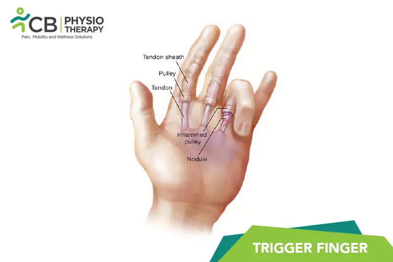
Understanding Trigger Finger
Trigger Finger (also called Stenosing Tenosynovitis) is one of the conditions our physiotherapists treat most often — and one of the most treatable when the actual driver is found. Two people with Trigger Finger rarely share the same underlying cause, which is why one-size-fits-all treatment so often disappoints.
What matters clinically is the pattern: how it started, what makes it better or worse, and how it behaves through the day. That pattern — mapped in a structured assessment — tells us which structures are involved and which treatment will actually move the needle.
Causes & risk factors of Trigger Finger
What commonly drives it — your assessment pins down which of these apply to you.
Joint stiffness and age-related change
Joints that move less start to hurt more; age-related wear is normal, but painful stiffness is not something to simply accept.
Weakness and deconditioning
When supporting muscles lose strength, load shifts to structures that were never meant to carry it.
Old injuries and compensations
A previous injury that never fully rehabilitated often leaves a movement pattern that overloads something else.
Sudden spikes in activity
A new sport, a new gym program, a long trek — tissue adapts to gradual change and protests when change is sudden.
Muscle strain and overload
A sudden lift, an awkward movement or simply more load than the tissues were prepared for — the most common trigger.
When physiotherapy is the right call
If any of these sound like you, an assessment is the right first step.
- ✓ You want to return to sport or work with a plan, not by trial and error
- ✓ Trigger Finger that keeps returning or hasn't settled within two weeks
- ✓ Pain or stiffness that limits work, sleep or daily activities
- ✓ You want to avoid long-term painkillers — or surgery has been mentioned
- ✓ You've been diagnosed with Trigger Finger and told to "rest and see"
Your treatment journey
Step by step — from your first assessment to a tracked recovery plan.
A detailed first assessment
History, movement testing and strength checks — a focused session to find what is actually driving your Trigger Finger.
A diagnosis you understand
Your physiotherapist explains the findings in plain language — what is affected, why it happened and what that means for you.
Active, tracked treatment
Hands-on therapy, the right modalities and graded exercise — progressed session by session and tracked on Fizo IQ™.
Prevention, built in
The final phase rebuilds strength and habits so the same problem does not come back — with a home plan you keep.
What recovery looks like
- ✓ Relief that comes from treating the cause, not masking it
- ✓ A clear diagnosis and a timeline you can plan around
- ✓ Strength and movement rebuilt, not just pain reduced
- ✓ Lower chance of recurrence, with a prevention plan
- ✓ Care without long-term medicines — at a clinic near you or at home
When to see a doctor — red flags
How CB Physiotherapy treats Trigger Finger
At CB Physiotherapy, treatment for Trigger Finger starts with finding its cause: a structured, physiotherapist-led assessment before anything is prescribed. Your plan then combines hands-on therapy, targeted exercise and the right modalities — delivered at a clinic near you or at home, and coordinated with your doctor where medical care is involved.
Every case runs on Fizo IQ™, our recovery engine: diagnosis, root cause, plan, milestones and goals, tracked session by session. You always know where you are in your recovery — and what comes next.
Get Trigger Finger treatment near you
Certified physiotherapists at a clinic near you or at home — same-day slots, assessment-led care.
Request received!
We'll call you within 10 minutes to confirm your home visit.We'll call you within 10 minutes to confirm your slot.
Chat with us on WhatsAppTrigger Finger, answered
1: Finger stiffness: Stiffness in the affected finger, especially in the morning or after periods of inactivity.
2: Finger locking or catching: The finger may get stuck in a bent position (flexed) and then suddenly straighten with a snapping or popping sensation.
3: Pain or tenderness: There may be pain or tenderness at the base of the affected finger, where the tendon is inflamed.
4: Swelling: You might notice swelling or a bump (nodule) at the base of the affected finger.
5: Difficulty straightening the finger: It may become challenging to fully extend the affected finger, and you may need to use your other hand to help straighten it.
6: Clicking sensation: You may feel a clicking or popping sensation when moving the affected finger.
1: Repetitive movements: Activities or occupations that involve repetitive gripping or grasping motions can strain the tendons in the fingers, leading to inflammation and triggering of the condition.
2: Medical conditions: Certain medical conditions, such as rheumatoid arthritis, diabetes, gout, or hypothyroidism, can increase the risk of developing trigger finger. These conditions may affect the tendons or increase inflammation in the body, contributing to the development of the condition.
3: Age and gender: Trigger finger is more common in women and in individuals aged 40 to 60 years old. Hormonal changes and age-related changes in tendon structure may play a role in its development.
4: Hand anatomy: Anatomical factors, such as the shape and size of the tendons or the presence of nodules or bumps on the tendons, can increase the likelihood of the tendon getting caught in the tendon sheath, leading to triggering.
5: Injury or trauma: Injuries to the hand or fingers, such as fractures or lacerations, can damage the tendons or tendon sheaths, increasing the risk of developing trigger finger.
6: Overuse or strain: Excessive use of the fingers or hands, particularly during activities that involve repetitive gripping or forceful hand movements, can strain the tendons and contribute to inflammation and triggering.
Pathology
The pathology of trigger finger involves changes in the affected tendon and its surrounding structures, particularly the tendon sheath. The condition usually starts with inflammation of the flexor tendon, which is tendon responsible for bending the affected finger. This inflammation can be due to repetitive use, injury, or underlying medical conditions such as arthritis. In some cases, nodules or bumps may develop on the flexor tendon. These nodules can further impede the tendon's movement within the tendon sheath and increase the likelihood of triggering.
1: Medical history assessment: Your healthcare provider will begin by asking about your symptoms, including any pain, stiffness, locking, or snapping sensations in your finger. They may inquire about your occupation, hobbies, or any recent injuries that could be contributing to your symptoms. Providing information about your medical history, including any underlying health conditions, can also help in the diagnostic process.
2: Physical examination: A physical examination is often performed to assess the affected finger and evaluate its range of motion. Your healthcare provider may manually manipulate the finger to feel for any nodules or bumps along the flexor tendon. They may also observe the triggering or snapping motion of the finger during movement. Additionally, they may check for signs of inflammation or tenderness at the base of the affected finger.
3: Provocative tests: During the physical examination, your healthcare provider may perform specific maneuvers to reproduce the triggering or snapping sensation in your finger. These provocative tests can help confirm the diagnosis of trigger finger and differentiate it from other conditions with similar symptoms.
4: Imaging studies: In some cases, imaging studies such as ultrasound or magnetic resonance imaging (MRI) may be recommended to further evaluate the affected finger and surrounding structures. Ultrasound can visualize the flexor tendon, tendon sheath, and any nodules or thickening present. MRI can provide detailed images of the soft tissues in the hand and may be useful in cases where the diagnosis is unclear or if there are concerns about other underlying conditions.
5: Electromyography (EMG): In rare instances, electromyography may be performed to assess the electrical activity of the muscles and nerves in the hand and forearm. EMG can help rule out nerve compression or other neurological conditions that may be contributing to finger symptoms.
Note: Medication should not be taken without the doctor’s prescription.
Surgery:
Surgery for trigger finger may be recommended if conservative treatments such as splinting, medication, or corticosteroid injections have not provided sufficient relief, or if the condition is severe and interfering with daily activities. The primary goal of surgery is to release the constricted tendon sheath, allowing the affected tendon to move more freely without catching or triggering. Here are the main surgical procedures used for trigger finger:
Percutaneous release: Also known as percutaneous trigger finger release or needle release, this minimally invasive procedure involves using a needle to release the tight tendon sheath. The surgeon inserts a needle into the base of the affected finger and uses it to release the constricted portion of the tendon sheath. Percutaneous release is typically performed under local anesthesia and can be done in an outpatient setting. It offers quicker recovery times and minimal scarring compared to traditional open surgery.
Open release surgery: In cases where percutaneous release is not feasible or effective, open release surgery may be performed. During this procedure, the surgeon makes a small incision at the base of the affected finger and carefully cuts and opens the constricted tendon sheath to create more space for the tendon to move freely. Open release surgery allows for direct visualization of the affected structures and may be preferred in cases of severe or complex trigger finger.
Endoscopic release: Endoscopic trigger finger release is a minimally invasive surgical technique that uses a thin, flexible tube with a camera (endoscope) to visualize and release the tight tendon sheath. The surgeon makes one or more small incisions near the base of the affected finger and inserts the endoscope and specialized surgical instruments to perform the release. Endoscopic release offers the advantages of smaller incisions, reduced trauma to surrounding tissues, and faster recovery times compared to open surgery.
Tenosynovectomy: In rare cases where trigger finger is associated with significant inflammation or scarring of the tendon sheath, a tenosynovectomy may be performed. This procedure involves removing the inflamed or scarred portion of the tendon sheath to relieve pressure on the affected tendon. Tenosynovectomy may be considered when other surgical options have been unsuccessful or when there are signs of more extensive tendon damage.
After surgery, patients typically undergo rehabilitation, which may include gentle exercises, splinting, and physical therapy to regain finger strength and mobility. Most individuals experience significant improvement in symptoms following surgical intervention for trigger finger, with a low risk of recurrence. However, as with any surgical procedure, there are potential risks and complications, and outcomes may vary depending on individual factors and the specific surgical technique used.
Custom splints or orthotic devices may be used to immobilize the affected finger in a neutral or extended position, particularly during periods of rest or sleep. Splinting can help reduce inflammation, prevent triggering, and promote proper alignment of the finger and tendon sheath. Physiotherapists can provide guidance on proper splinting techniques and monitor progress throughout the treatment process.
Ultrasound therapy:
Therapeutic ultrasound involves the use of high-frequency sound waves to generate heat deep within the tissues. Ultrasound therapy can help improve blood circulation, reduce inflammation, and promote tissue healing in the affected finger. Physiotherapists may use ultrasound in conjunction with other treatments such as manual therapy or exercise to enhance the effectiveness of therapy.
Electrical stimulation:
Electrical stimulation, also known as neuromuscular electrical stimulation (NMES) or transcutaneous electrical nerve stimulation (TENS), involves the application of electrical currents to the skin using electrodes. Electrical stimulation can help relieve pain, reduce muscle spasms, and improve muscle strength and function in the affected hand and finger. It may be used as part of a comprehensive rehabilitation program for trigger finger.
Interferential current therapy (IFC):
IFC is a form of electrical stimulation that uses two medium-frequency alternating currents to target deep tissues. It can help alleviate pain, reduce swelling, and improve blood flow in the affected finger. Physiotherapists may use IFC in combination with other modalities or manual techniques to address trigger finger symptoms.
Pulsed electromagnetic field therapy (PEMF):
PEMF therapy involves the use of electromagnetic fields to stimulate tissue repair and regeneration. It can help reduce pain and inflammation, improve circulation, and enhance healing in the affected finger. PEMF devices may be used by physiotherapists as part of a comprehensive treatment plan for trigger finger.
Iontophoresis:
Iontophoresis is a non-invasive technique that involves the use of a mild electrical current to deliver medication, typically a corticosteroid or anti-inflammatory agent, through the skin and into the affected tissues. Iontophoresis can help reduce inflammation and pain in the affected finger and may be used as an adjunctive treatment for trigger finger.
Manual therapy:
Hands-on techniques such as joint mobilizations, soft tissue mobilization, and massage may be used by physiotherapists to improve finger mobility, reduce muscle tension, and alleviate pain in the affected finger and hand.
Therapeutic exercises:
Physiotherapists may prescribe specific exercises to strengthen the muscles of the hand and forearm, improve finger flexibility and range of motion, and enhance overall hand function. These exercises may include stretching, grip strengthening exercises, tendon gliding exercises, and dexterity drills tailored to the individual's needs and abilities.
Functional training:
Functional activities and tasks relevant to the individual's daily life and occupation may be incorporated into the rehabilitation program to improve hand and finger function, coordination, and independence in performing activities of daily living.
Don't let Trigger Finger decide your day.
Get assessed today — find the cause, get a plan, and know when you'll feel better.