
Myositis Ossification (mo) — Symptoms, Causes & Treatment
Also known as Myositis Ossificans Traumatica. Causes, symptoms and how physiotherapy treats Myositis Ossification (mo) — without surgery or medicines.
Understanding Myositis Ossification (mo)
Myositis Ossification (mo) (also called Myositis Ossificans Traumatica) affects people differently, and the right care starts with understanding how it affects you. Its day-to-day impact — on movement, strength, energy and confidence — is where physiotherapy does its work.
Our role in Myositis Ossification (mo) is practical: protect and rebuild movement and daily function with a plan set after a careful assessment, in coordination with your doctor wherever medical management is involved. No two plans look the same, because no two cases behave the same.
Causes & risk factors of Myositis Ossification (mo)
What commonly drives it — your assessment pins down which of these apply to you.
More than one possible driver
Myositis Ossification (mo) can have several contributing factors — a careful history and physical assessment separate them rather than guessing.
Reduced movement and deconditioning
Whatever the trigger, less movement quickly adds weakness and stiffness of its own — usually the most reversible part.
Daily-life load and habits
Work posture, repeated tasks, sleep and stress all shape how a condition behaves day to day.
General health and history
Overall health, previous injuries and existing conditions influence both the picture and the plan — the assessment takes all of it in.
When physiotherapy is the right call
If any of these sound like you, an assessment is the right first step.
- ✓ A new diagnosis of Myositis Ossification (mo) and you want a clear rehabilitation start point
- ✓ Daily tasks, walking or stairs are getting harder than they used to be
- ✓ Strength, stamina or balance has visibly dropped
- ✓ You want a structured clinic + home program with measurable milestones
- ✓ Family members need guidance to support you safely
Your treatment journey
Step by step — from your first assessment to a tracked recovery plan.
A detailed first assessment
History, movement testing and strength checks — a focused session to find what is actually driving your Myositis Ossification (mo).
A diagnosis you understand
Your physiotherapist explains the findings in plain language — what is affected, why it happened and what that means for you.
Active, tracked treatment
Hands-on therapy, the right modalities and graded exercise — progressed session by session and tracked on Fizo IQ™.
Prevention, built in
The final phase rebuilds strength and habits so the same problem does not come back — with a home plan you keep.
What recovery looks like
- ✓ More independence in daily tasks
- ✓ Better strength, balance and stamina
- ✓ Fewer setbacks, with red flags monitored
- ✓ A program your family understands and can support
- ✓ Progress measured milestone by milestone on Fizo IQ™
When to see a doctor — red flags
How CB Physiotherapy treats Myositis Ossification (mo)
We treat Myositis Ossification (mo) the way we treat everything: assessment first, plan second, treatment third. It sounds obvious, but it is the difference between sessions that fill a slot and sessions that move you towards a goal. Care is delivered by certified physiotherapists at a clinic near you or at home.
Your whole journey is tracked on Fizo IQ™ — the diagnosis, the root cause, the milestones and the goals — so progress is measured, not guessed, and the plan adapts the moment your response calls for it.
Get Myositis Ossification (mo) treatment near you
Certified physiotherapists at a clinic near you or at home — same-day slots, assessment-led care.
Request received!
We'll call you within 10 minutes to confirm your home visit.We'll call you within 10 minutes to confirm your slot.
Chat with us on WhatsAppMyositis Ossification (mo), answered
1: Blunt Trauma: Direct impact injuries, such as muscle contusions from contact sports or accidents, are the most common causes.
2: Repetitive Microtrauma: Recurrent minor injuries or strains from repetitive motion (e.g., in athletes or individuals with physically demanding jobs) can trigger the condition.
3: Muscle Strains or Tears: Significant muscle injuries involving partial or complete tears can lead to ossification during the healing process.
4: Surgical Procedures: Postoperative complications from surgeries involving muscles or soft tissues may contribute to developing Myositis Ossification.
5: Severe Fractures: Bone fractures that extend into muscle tissue can cause abnormal bone formation within the muscle during the healing phase.
6: Neurological Conditions: In some cases, individuals with spinal cord injuries or traumatic brain injuries may develop Myositis Ossification without direct muscle trauma.
7: Genetic Factors: Rarely, hereditary conditions like Fibrodysplasia Ossificans Progressiva can cause widespread ossification, although this is different from trauma-induced Myositis Ossification.
8: Idiopathic Cases: Occasionally, Myositis Ossification can occur without a clear cause, although this is less common.
1: Pain and Tenderness: Persistent or worsening pain at the site of injury.
2: Swelling: Noticeable swelling or inflammation in the affected area.
3: Hard Lump Formation: The presence of a hard, bony lump within the muscle.
4: Reduced Range of Motion: Difficulty moving the affected limb or joint due to stiffness.
5: Warmth and Redness: The area may feel warm and look red initially, similar to an inflammatory response.
6: Muscle Weakness: Reduced strength in the muscle near the affected area.
7: Bruising: In cases following trauma, bruising might be present alongside swelling.
These symptoms typically develop a few weeks after the initial trauma and can progress over time as the ossification process continues.
Pathology
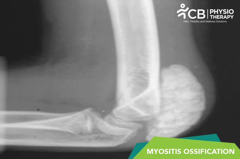
Myositis Ossification involves the abnormal formation of bone tissue within muscle following trauma. Initially, the injury triggers an inflammatory response, leading to fibroblastic proliferation. Over time, these fibroblasts differentiate into osteoblasts, resulting in the deposition of bone matrix within the soft tissue. This ectopic bone formation matures in a zonal pattern, with a central, less organized core and a more mature, ossified outer layer. The process usually stabilizes over weeks to months, and the bone becomes fully formed within 6-12 months.
Initial assessment based on patient history, symptoms, and physical examination to identify signs such as swelling, pain, and a palpable lump.
X-ray Imaging:
X-ray Imaging can reveal ossification typically 2-3 weeks after injury. It shows characteristic zonal ossification with mature bone at the periphery and less mature tissue centrally.
Ultrasound:
Ultrasound is useful for early detection when X-rays may not show changes. It can help identify soft tissue changes and the early presence of calcification.
Magnetic Resonance Imaging (MRI):
MRI helps detect soft tissue inflammation and edema in the early phase. It helps differentiate Myositis Ossification from malignant tumors.
Computed Tomography (CT) Scan:
CT scans help provide detailed imaging of bone formation and the extent of ossification. It is useful for surgical planning if removal is necessary.
Bone Scan (Scintigraphy):
Bone Scan demonstrates increased metabolic activity in the area of ossification. It is primarily used for confirmation in complex cases.
(Note: Medication should not be taken without the doctor’s prescription.)
Surgery:
1: Excision of the Ossified Mass:
Complete Surgical Resection: The abnormal bone is removed to restore function and reduce pain.
Typically performed after the ossification has matured (usually 6-12 months post-injury) to minimize the risk of recurrence.
2: Minimally Invasive Techniques:
In some cases, arthroscopic or smaller incisions may be used for targeted removal, reducing recovery time.
3: Joint Preservation Surgery:
If the ossification affects a joint, surgery may include techniques to preserve or restore joint function while removing the bone.
4: Rehabilitation Post-Surgery:
Postoperative physiotherapy is essential to restore range of motion, strengthen muscles, and prevent stiffness.
Thermotherapy/ Cryotherapy:
Thermotherapy (heat therapy) and cryotherapy (cold therapy) help to reduce pain and inflammation.
Ultrasound Therapy:
1: Uses high-frequency sound waves to promote tissue healing and reduce inflammation.
2: May help improve blood flow to the area, supporting pain relief and flexibility.
Transcutaneous Electrical Nerve Stimulation (TENS):
1: Provides pain relief by delivering low-voltage electrical currents through the skin.
2: Effective for managing pain without the use of medication.
Interferential Current Therapy (IFC):
1: Uses two medium-frequency currents that intersect to create a low-frequency effect at the treatment site.
2: Helps reduce pain, swelling, and muscle spasm.
Pulsed Shortwave Diathermy:
1: Uses electromagnetic waves to deliver deep heat to tissues.
2: Can help decrease stiffness, increase blood circulation, and reduce pain.
Laser Therapy (Low-Level Laser Therapy):
1: Uses light energy to promote tissue healing and reduce pain and inflammation.
2: Can be used as part of a comprehensive pain management plan.
Range of Motion (ROM) Exercises
1: Early Stage: Gentle, pain-free ROM exercises to prevent stiffness and maintain joint mobility.
2: Progressive Stretching: As tolerated, more active and passive stretching exercises are introduced to prevent loss of function and joint stiffness.
Strengthening Exercises
1: Isometric Exercises: Start with static muscle contractions to strengthen muscles without moving the affected joint.
2: Progressive Resistance Training: Introduced once pain and swelling are under control to rebuild muscle strength.
Functional Training
1: Task-Specific Movements: Training aimed at regaining the ability to perform daily activities.
2: Gait Training: For MO affecting lower extremities, gait training may be incorporated to improve walking patterns and reduce compensatory movements.
Manual Therapy
1: Soft Tissue Mobilization: Gentle techniques to reduce muscle tension and improve circulation around the affected area.
2: Joint Mobilizations: If safe, mobilizations can help restore normal joint movement.
Don't let Myositis Ossification (mo) decide your day.
Get assessed today — find the cause, get a plan, and know when you'll feel better.