Open today · 8 AM–8 PM · Same-day slots
CB Physiotherapy
Same-day appointments

Myositis Ossification (mo) Treatment Doctors in Ghaziabad

Whether it's a dull ache, a sharp catch, or pain that keeps coming back — we find the root cause of your Myositis Ossification (mo) and fix it, at a clinic near you or at home.

Precision Care, Proven Recovery
ACCURATE DIAGNOSIS
ROOT CAUSE ASSESSMENT
COMPREHENSIVE RECOVERY PLAN
MILESTONE TRACKING
GOALS ACHIEVED
STAYING ACTIVE
See how it works ↓
Move pain-free Avoid drugs & surgery
5.0 / 5 · 20 Google reviews Verified on Google

Request received!

We'll call you within 10 minutes to confirm your home visit.We'll call you within 10 minutes to confirm your slot.

Chat with us on WhatsApp

Book in 30 seconds

Visit your nearest CB clinic. Tell us your concern and a preferred time — we'll confirm your slot by call within 10 minutes.

Same-day booking by default. We'll confirm your exact slot by call.
+91
No spam · Your number is safe with us
Prefer to talk now? Call +09821966696
Understanding your myositis ossification (mo)

Does this sound like you?

Common triggers include muscle strain, joint stiffness, poor posture and long desk hours. If two or more of these fit, a physiotherapy assessment can identify exactly what's driving it.

  • A dull ache or sharp, burning myositis ossification (mo) that keeps returning
  • Discomfort that worsens with movement, lifting or long sitting hours
  • Morning stiffness that eases as you move
  • Pain that limits work, sport or everyday activities
  • Recurring episodes that keep coming back

Most myositis ossification (mo) improves without surgery or long-term medication — the key is finding the cause first.

⚠ When to seek urgent medical care

Myositis Ossification (mo) rarely signals something serious, but see a doctor immediately if it comes with:

  • Fever, unexplained weight loss, or a history of cancer
  • Numbness or weakness that keeps getting worse
  • Loss of bladder or bowel control
  • Pain after a major fall or accident

Our physios screen for these red flags at every first assessment and refer you to the right doctor if needed.

Assessment-led treatment

How we treat myositis ossification (mo)

We start with an AI-based assessment to find the root cause — then follow a clear, four-phase plan.

Phase 1

Ease the pain

Hands-on therapy and advanced modalities to calm pain and inflammation fast.

Phase 2

Restore movement

Mobility work and posture correction so everyday movement feels normal again.

Phase 3

Build strength

Targeted strengthening that protects you — in-clinic or in our physio gym.

Phase 4

Return to activity

Back to work, sport and life — with a home program that prevents relapse.

Your physiotherapist, with Fizo IQ™

Every recovery, planned and proven

One patient record, stage by stage: your physiotherapist pinpoints the diagnosis, finds the root cause and builds your in-clinic plan — while Fizo IQ™, our AI decision-support engine, tracks every milestone until your goals are met and you leave with an exercise plan to stay active.

Physio Expert @CB with Fizo IQ™ — recovery engine
TRACKING
ACCURATE DIAGNOSIS
L4–L5 disc bulge · nerve-root irritation, pinpointed at the first movement assessment
ROOT CAUSE ASSESSMENT
Poor sitting posture, weak core–hip chain · quietly loading that disc
COMPREHENSIVE RECOVERY PLAN
24 clinic sessions · manual therapy + spinal decompression · guided exercise therapy, reviewed weekly
MILESTONE TRACKING
Sitting pain-free ✓ · stairs ✓ · next: the morning 5 km walk
GOALS ACHIEVED
Pain 7 → 3 in six weeks · back to daily life, moving freely
STAYING ACTIVE
A personalized exercise plan, yours to keep · so the pain doesn't come back
Example recovery journey · every plan is built around you
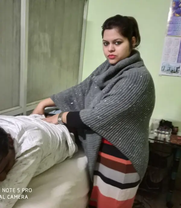
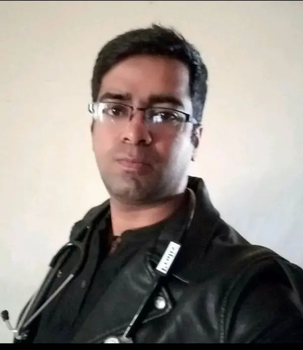
Meet your care team

Licensed experts who treat hundreds like you every month

Every CB physio is a qualified BPT/MPT clinician. You don't need to choose one — our lead physiotherapist reviews your case and assigns the specialist best suited to treat it.

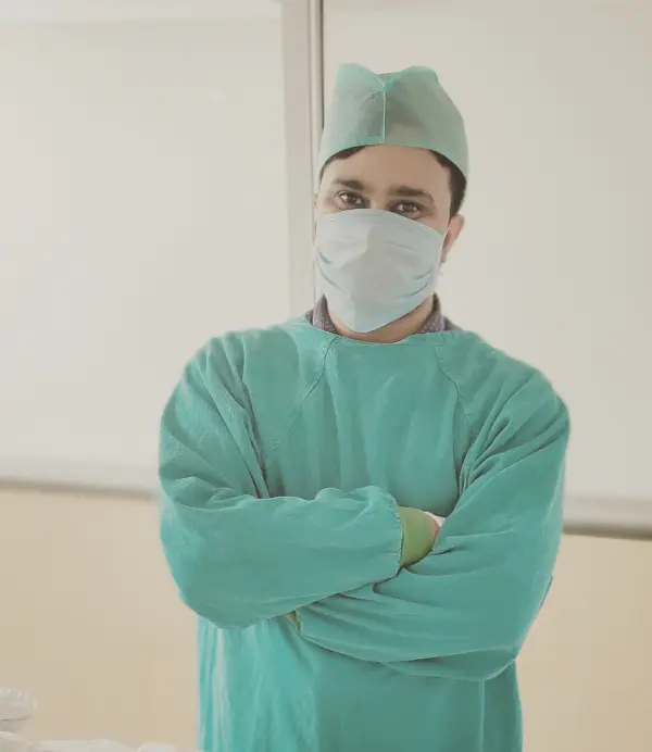
Dr. Puneet Kumar, physiotherapist in Ghaziabad Verified

Dr. Puneet Kumar

Physiotherapist · MPT (Orthopaedics)

5.0 (1)
Ghaziabad
6 yrs experience
Ortho Spine Sports Physiotherapy Center
Not sure who to see? You don't have to decide. Our lead physiotherapist reviews every case and assigns the physio best suited to it — based on your condition and availability.
Patient stories

Myositis Ossification (mo) recoveries, verified on Google

Rated 5.0 / 5 across 20 verified patient reviews in Ghaziabad.

Verified

“मुझे फिजियोथेरेपी करवानी है होम सर्विस आपके पास अवेलेबल है”

SK
Sumit Kumar Verified patient · Treated at CB · 5 months ago
Verified

“VERY Best physiotherapist and good experience”

AU
Arun Upadhyay Verified patient · Treated at CB
Verified

“I recently had the pleasure of receiving physiotherapy treatment from Dr. Asif, and I cannot praise his skills and dedication enough. As a physiotherapist, he possesses a unique blend of expertise, compassion, and professionalism that sets him apart from others in his field.”

SK
Shawaiz Khan Verified patient · Treated at CB · 3 months ago
Verified

“I recently had the privilege of being treated by Dr. Asif Alam, and I cannot speak highly enough of his exceptional care and expertise as a physiotherapist. His ability to diagnose and explain the root causes of my issues was truly impressive. He provided me with a clear understanding of my condition His hands-on techniques were precise and effective, providing me with immediate relief and tangible results. He continuously monitored my progress and adjusted the treatment plan accordingly, ensuring that I was on track to a full recovery.”

KN
Krishna N12 Verified patient · Treated at CB · 3 months ago
Verified

“I have no words to express how good Dr. Asif was for post-knee rehabilitation. Every session I had was excellent. Day by day, I'm improving. I can walk without support. Thanks so much! All praise for you!🙏”

RB
Rajiv Bhatia Verified patient · Treated at CB · 3 months ago
Verified

“Thank you, Dr. shally for your outstanding service!👍🏼”

AB
Aakash Bansal Verified patient · Treated at CB · 4 months ago
Good to know

Myositis Ossification (mo), answered

Myositis Ossification (MO) is a condition characterized by the abnormal formation of bone tissue inside muscle or soft tissue after an injury. It typically develops after trauma, such as a muscle strain, contusion, or repetitive injury, and can lead to pain, stiffness, and a decrease in the range of motion of the affected area.
Myositis Ossification typically occurs after a trauma or certain conditions that lead to the abnormal formation of bone within a muscle or soft tissue. Here are the main causes:

1: Blunt Trauma: Direct impact injuries, such as muscle contusions from contact sports or accidents, are the most common causes.
2: Repetitive Microtrauma: Recurrent minor injuries or strains from repetitive motion (e.g., in athletes or individuals with physically demanding jobs) can trigger the condition.
3: Muscle Strains or Tears: Significant muscle injuries involving partial or complete tears can lead to ossification during the healing process.
4: Surgical Procedures: Postoperative complications from surgeries involving muscles or soft tissues may contribute to developing Myositis Ossification.
5: Severe Fractures: Bone fractures that extend into muscle tissue can cause abnormal bone formation within the muscle during the healing phase.
6: Neurological Conditions: In some cases, individuals with spinal cord injuries or traumatic brain injuries may develop Myositis Ossification without direct muscle trauma.
7: Genetic Factors: Rarely, hereditary conditions like Fibrodysplasia Ossificans Progressiva can cause widespread ossification, although this is different from trauma-induced Myositis Ossification.
8: Idiopathic Cases: Occasionally, Myositis Ossification can occur without a clear cause, although this is less common.

The symptoms of Myositis Ossification can vary depending on the severity and location of the condition but generally include:

1: Pain and Tenderness: Persistent or worsening pain at the site of injury.
2: Swelling: Noticeable swelling or inflammation in the affected area.
3: Hard Lump Formation: The presence of a hard, bony lump within the muscle.
4: Reduced Range of Motion: Difficulty moving the affected limb or joint due to stiffness.
5: Warmth and Redness: The area may feel warm and look red initially, similar to an inflammatory response.
6: Muscle Weakness: Reduced strength in the muscle near the affected area.
7: Bruising: In cases following trauma, bruising might be present alongside swelling.

These symptoms typically develop a few weeks after the initial trauma and can progress over time as the ossification process continues.

Pathology
Myositis Ossification involves the abnormal formation of bone tissue within muscle following trauma. Initially, the injury triggers an inflammatory response, leading to fibroblastic proliferation. Over time, these fibroblasts differentiate into osteoblasts, resulting in the deposition of bone matrix within the soft tissue. This ectopic bone formation matures in a zonal pattern, with a central, less organized core and a more mature, ossified outer layer. The process usually stabilizes over weeks to months, and the bone becomes fully formed within 6-12 months.
Clinical Evaluation:
Initial assessment based on patient history, symptoms, and physical examination to identify signs such as swelling, pain, and a palpable lump.

X-ray Imaging:
X-ray Imaging can reveal ossification typically 2-3 weeks after injury. It shows characteristic zonal ossification with mature bone at the periphery and less mature tissue centrally.

Ultrasound:
Ultrasound is useful for early detection when X-rays may not show changes. It can help identify soft tissue changes and the early presence of calcification.

Magnetic Resonance Imaging (MRI):
MRI helps detect soft tissue inflammation and edema in the early phase. It helps differentiate Myositis Ossification from malignant tumors.

Computed Tomography (CT) Scan:
CT scans help provide detailed imaging of bone formation and the extent of ossification. It is useful for surgical planning if removal is necessary.

Bone Scan (Scintigraphy):
Bone Scan demonstrates increased metabolic activity in the area of ossification. It is primarily used for confirmation in complex cases.
Medication: Nonsteroidal Anti-Inflammatory Drugs (NSAIDs), Pain Relievers, Bisphosphonates, Corticosteroids, Muscle Relaxants, Anticoagulants, etc.
(Note: Medication should not be taken without the doctor’s prescription.)

Surgery:

1: Excision of the Ossified Mass:
Complete Surgical Resection: The abnormal bone is removed to restore function and reduce pain.
Typically performed after the ossification has matured (usually 6-12 months post-injury) to minimize the risk of recurrence.

2: Minimally Invasive Techniques:
In some cases, arthroscopic or smaller incisions may be used for targeted removal, reducing recovery time.

3: Joint Preservation Surgery:
If the ossification affects a joint, surgery may include techniques to preserve or restore joint function while removing the bone.

4: Rehabilitation Post-Surgery:
Postoperative physiotherapy is essential to restore range of motion, strengthen muscles, and prevent stiffness.

Thermotherapy/ Cryotherapy:
Thermotherapy (heat therapy) and cryotherapy (cold therapy) help to reduce pain and inflammation.

Ultrasound Therapy:
1: Uses high-frequency sound waves to promote tissue healing and reduce inflammation.
2: May help improve blood flow to the area, supporting pain relief and flexibility.


Transcutaneous Electrical Nerve Stimulation (TENS):
1: Provides pain relief by delivering low-voltage electrical currents through the skin.
2: Effective for managing pain without the use of medication.


Interferential Current Therapy (IFC):
1: Uses two medium-frequency currents that intersect to create a low-frequency effect at the treatment site.
2: Helps reduce pain, swelling, and muscle spasm.


Pulsed Shortwave Diathermy:
1: Uses electromagnetic waves to deliver deep heat to tissues.
2: Can help decrease stiffness, increase blood circulation, and reduce pain.


Laser Therapy (Low-Level Laser Therapy):
1: Uses light energy to promote tissue healing and reduce pain and inflammation.
2: Can be used as part of a comprehensive pain management plan.


Range of Motion (ROM) Exercises
1: Early Stage: Gentle, pain-free ROM exercises to prevent stiffness and maintain joint mobility.
2: Progressive Stretching: As tolerated, more active and passive stretching exercises are introduced to prevent loss of function and joint stiffness.

Strengthening Exercises
1: Isometric Exercises: Start with static muscle contractions to strengthen muscles without moving the affected joint.
2: Progressive Resistance Training: Introduced once pain and swelling are under control to rebuild muscle strength.

Functional Training
1: Task-Specific Movements: Training aimed at regaining the ability to perform daily activities.
2: Gait Training: For MO affecting lower extremities, gait training may be incorporated to improve walking patterns and reduce compensatory movements.

Manual Therapy
1: Soft Tissue Mobilization: Gentle techniques to reduce muscle tension and improve circulation around the affected area.
2: Joint Mobilizations: If safe, mobilizations can help restore normal joint movement.

The patient is guided on modifying activities to avoid aggravating the condition. Educating the patient about tailored exercises for continued improvement outside the clinic helps maximize mobility and function while preventing further complications, making it a vital part of the management plan for Myositis Ossification.
Ready when you are

Don't let the pain decide your day.

Get assessed today — find the cause, get a plan, and know when you'll feel better.

📞 Call 💬 WhatsApp Book now