
Patella Dislocation — Symptoms, Causes & Treatment
Also known as Patella Dislocation. Causes, symptoms and how physiotherapy treats Patella Dislocation — without surgery or medicines.
Understanding Patella Dislocation
Patella Dislocation is one of the conditions our physiotherapists treat most often — and one of the most treatable when the actual driver is found. Two people with Patella Dislocation rarely share the same underlying cause, which is why one-size-fits-all treatment so often disappoints.
What matters clinically is the pattern: how it started, what makes it better or worse, and how it behaves through the day. That pattern — mapped in a structured assessment — tells us which structures are involved and which treatment will actually move the needle.
Causes & risk factors of Patella Dislocation
What commonly drives it — your assessment pins down which of these apply to you.
Old injuries and compensations
A previous injury that never fully rehabilitated often leaves a movement pattern that overloads something else.
Sudden spikes in activity
A new sport, a new gym program, a long trek — tissue adapts to gradual change and protests when change is sudden.
Muscle strain and overload
A sudden lift, an awkward movement or simply more load than the tissues were prepared for — the most common trigger.
Posture and long static hours
Desks, driving and screens keep the same structures loaded for hours at a stretch — comfortable at first, aching later.
Joint stiffness and age-related change
Joints that move less start to hurt more; age-related wear is normal, but painful stiffness is not something to simply accept.
When physiotherapy is the right call
If any of these sound like you, an assessment is the right first step.
- ✓ You've been diagnosed with Patella Dislocation and told to "rest and see"
- ✓ Movement feels restricted, weak or guarded compared to before
- ✓ You want to return to sport or work with a plan, not by trial and error
- ✓ Patella Dislocation that keeps returning or hasn't settled within two weeks
- ✓ Pain or stiffness that limits work, sleep or daily activities
Your treatment journey
Step by step — from your first assessment to a tracked recovery plan.
A detailed first assessment
History, movement testing and strength checks — a focused session to find what is actually driving your Patella Dislocation.
A diagnosis you understand
Your physiotherapist explains the findings in plain language — what is affected, why it happened and what that means for you.
Active, tracked treatment
Hands-on therapy, the right modalities and graded exercise — progressed session by session and tracked on Fizo IQ™.
Prevention, built in
The final phase rebuilds strength and habits so the same problem does not come back — with a home plan you keep.
What recovery looks like
- ✓ A clear diagnosis and a timeline you can plan around
- ✓ Strength and movement rebuilt, not just pain reduced
- ✓ Lower chance of recurrence, with a prevention plan
- ✓ Care without long-term medicines — at a clinic near you or at home
- ✓ Relief that comes from treating the cause, not masking it
When to see a doctor — red flags
How CB Physiotherapy treats Patella Dislocation
At CB Physiotherapy, treatment for Patella Dislocation starts with finding its cause: a structured, physiotherapist-led assessment before anything is prescribed. Your plan then combines hands-on therapy, targeted exercise and the right modalities — delivered at a clinic near you or at home, and coordinated with your doctor where medical care is involved.
Every case runs on Fizo IQ™, our recovery engine: diagnosis, root cause, plan, milestones and goals, tracked session by session. You always know where you are in your recovery — and what comes next.
Exercises that help with Patella Dislocation
Make these exercises yours — a CB physio personalizes, doses and tracks your plan week by week.
Get my personalized planGeneral guidance only — stop if an exercise increases your pain, and check with your physio before starting a new routine.
Get Patella Dislocation treatment near you
Certified physiotherapists at a clinic near you or at home — same-day slots, assessment-led care.
Request received!
We'll call you within 10 minutes to confirm your home visit.We'll call you within 10 minutes to confirm your slot.
Chat with us on WhatsAppPatella Dislocation, answered
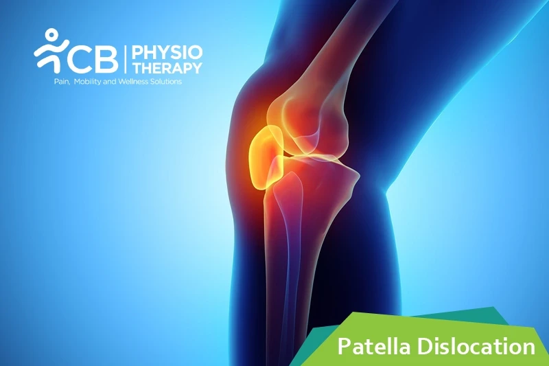
Patella dislocation is a dislocation of the kneecap i.e the patella from its groove at the knee joint. The knee joint is a joint at which three bones meet the thighbone, the shinbone, and the patella. Normally, when the knee is bent and straightened, the kneecap slides up and down inside a groove, which is between the bottom end of the thighbone and the upper end of the shinbone, this groove is known as the trochlear groove. When the patella dislocates, it's forced out of the trochlear groove and can no longer move up and down. This locks the knee pulls the ligaments (that secures the knee joint) out of place and often tears them. Patella dislocation is an acute injury caused by force. It is a common injury since the kneecap takes less force to dislocate.
Patellar dislocation can occur due to many reasons, a few of them are mentioned below:
- Due to direct or an indirect force,
- Fall from a height,
- A collision that knocks the kneecap out of place,
- Sudden turn that twists the knee while the lower leg is still firmly planted,
- Ligamentous laxity,
- Muscle imbalance like weakness of the vastus medialis obligue,
- High impact sports, like athletes,
- Previous injury or by another preexisting anatomical condition,
- Congenital patellar dislocation (trochlear dysplasia) developmental abnormalities like cerebral palsy, down syndrome, patellar Alta, etc,
- Obese people, whose joints are under more pressure,
- People with patellar instability, especially if they have already dislocated their patella.
It depends on how far the patellar bone has been forced out of the knee joint, and to what extent the surrounding muscles and ligaments, blood vessels, and nerves have been injured. The symptoms include:
- An audible pop,
- Buckling of the knee,
- Intense pain and tenderness,
- Sudden swelling,
- Bruising at the knee,
- Locking of the knee,
- Inability to walk.
- Kneecap visually out of place,
- Deformed knee,
- The knee is bent and cannot be straightened out,
- Hypermobile patella.
PATHOLOGY
Patella dislocation is influenced by minor alterations or derangements factors. The sulcus angle should not be more than 138 degrees and the Q angle should not exceed15 degrees. Both patella Alta and the weakness and atrophy of the vastus medialis obliquus, influences the dislocation tendency and may cause muscular imbalance.
Physical examination:
Physical examination of the knee is done thoroughly to check the dislocation, joint effusion, and bleeding. The patient is assessed for femoral anteversion, tibial torsion, patellar Alta, genu recurvatum, genu valgus, genu varum, etc.
X-ray:
X-ray helps to show dislocation, fracture, malalignment, loose bodies, arthritic changes, it also helps to exclude associated fractures like osteochondral, avulsion, etc.
Computed Tomography:
Computed Tomography helps to measure the distance between tibial tuberosity and trochlear groove.
Magnetic resonance imaging (MRI):
Magnetic resonance imaging (MRI) is a gold standard and helps to check dislocation and the degree of tear.
Medication: NSAID's, pain killers, muscle relaxants, etc.
Note: Medication should not be taken without a doctor's prescription.
Reduction:
Reduction is a process of manually relocating the kneecap.
Surgery: In case of damage to the bone or the cartilage and tendons of the knee, surgery is recommended. Types of surgeries include:
- Arthroscopy
- Lateral release
- Medial patellofemoral ligament reattachment
- Reconstruction
- Osteotomy
- Trocleoplasty
Elevation:
The affected limb is kept in an elevated position to help keep the swelling down.
Bracing:
Bracing is done to help control the position of the patella by preventing dislocation but care should be taken that the brace should not be overused.
Cryotherapy helps to decrease swelling and pain.
Thermotherapy is used for relaxation of the muscles which in turn helps to perform the exercise program.
Ultrasound is very effective in breaking adhesions and improving flexibility.
Transcutaneous electrical nerve stimulation (TENS):
Transcutaneous electrical nerve stimulation provides relief from pain, which may occur after performing stretching exercises.
Laser therapy is used for the pain management of patients having dislocation of the patella.
Kinesio-taping helps reduce the movement of the patella, it holds the patella in the proper position while the muscles around the knee work properly to hold the patella in position.
Manual therapy consists of different techniques like manipulation, joint mobilization, and soft tissue mobilization. It helps in decreasing pain and improves functional mobility of the knee joint.
Range of motion exercises:
Range of motion exercises help to improve the contraction of the knee muscles. These exercises are very important to improve the flexibility of the muscles while the joint is restabilized. Pelvic stabilization exercises help strengthen the hip flexors and hip abductors, which helps to stabilize the function of the extremity.
Strengthening exercises like quadriceps, hamstrings, adductors, hip, and lower abdomen are recommended. Closed kinetic chain exercises are also done. Advanced hip strengthening is done after a few weeks to help the patient prepare to return to normal activity.
Stretching exercises:
Stretching exercises improve the flexibility of hamstrings and quadriceps. These exercises are essential to ensure that each muscle group is offering the full range of mobility.
Proprioception and balance exercises:
Proprioceptive and balance exercises are also incorporated in the exercise program to improve the stability and balance of the knee.
Gait training:
Gait training is given gradually with crutches and a brace to hold the joint in place.
The patient is advised to continue doing the exercises for a lifelong. If he/ she is an athlete, then he/ she needs to practice vigorous movements and exercises and should be done properly. Incorrectly doing the exercises can put repetitive stress on the muscles and joints.
Don't let Patella Dislocation decide your day.
Get assessed today — find the cause, get a plan, and know when you'll feel better.